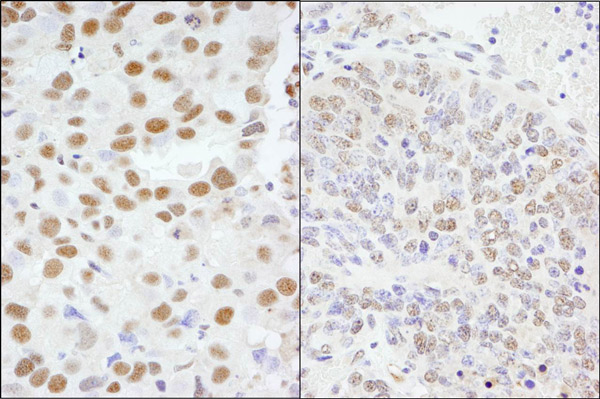
Immunohistochemistry (Formalin/PFA-fixed paraffin-embedded sections) analysis of human breast carcinoma (left) and mouse teratoma (right) tissues labelling AKAP8 with ab72196 at 1/1000 (0.2µg/ml) and 1/200 (1µg/ml). Detection: DAB.
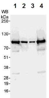
All lanes : Anti-AKAP8 antibody (ab72196) at 0.04 µg/mlLane 1 : HeLa whole cell lysate at 50 µgLane 2 : HeLa whole cell lysate at 15 µgLane 3 : HeLa whole cell lysate at 5 µgLane 4 : 293T whole cell lysate at 50 µgdeveloped using the ECL technique
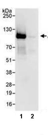
Immunoprecipitation of HeLa whole cell lysate (1 mg) using ab72196 at 3 µg/mg lysate. 20% of the immunoprecipitate was used for Western blot and bands were detected using ab72196 at 1 µg/ml. Bands were developed using chemiluminescence with an exposure time of 3 seconds. Lane 2 represents a Control IgG.