WDR82-Antibody-N-term
| Name | WDR82-Antibody-N-term |
|---|---|
| Supplier | Abgent, a WuXi AppTec company |
| Catalog | AP20978b |
| Prices | $99.00, $295.00 |
| Sizes | 80 µl, 400 µl |
| Host | Rabbit |
| Clonality | Polyclonal |
| Isotype | Rabbit Ig |
| Applications | WB ICC/IF ELISA |
| Species Reactivities | Human |
| Purity/Format | Purified polyclonal antibody supplied in PBS with 0.09% (W/V) sodium azide. This antibody is purified through a protein A column, followed by peptide affinity purification. |
| Description | Rabbit Polyclonal |
| Gene | WDR82 |
| Supplier Page | Shop |
Product images
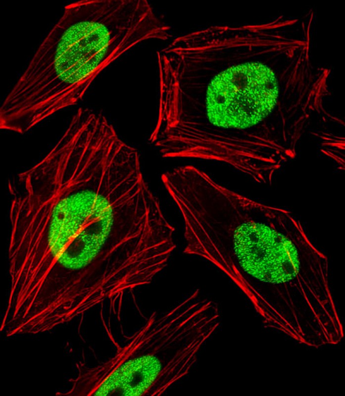 Fluorescent image of Hela cells stained with WDR82 Antibody (N-term)(Cat#AP20978b). AP20978b was diluted at 1:25 dilution. An Alexa Fluor 488-conjugated goat anti-rabbit lgG at 1:400 dilution was used as the secondary antibody (green). Cytoplasmic actin was counterstained with Alexa Fluor® 555 conjugated with Phalloidin (red).
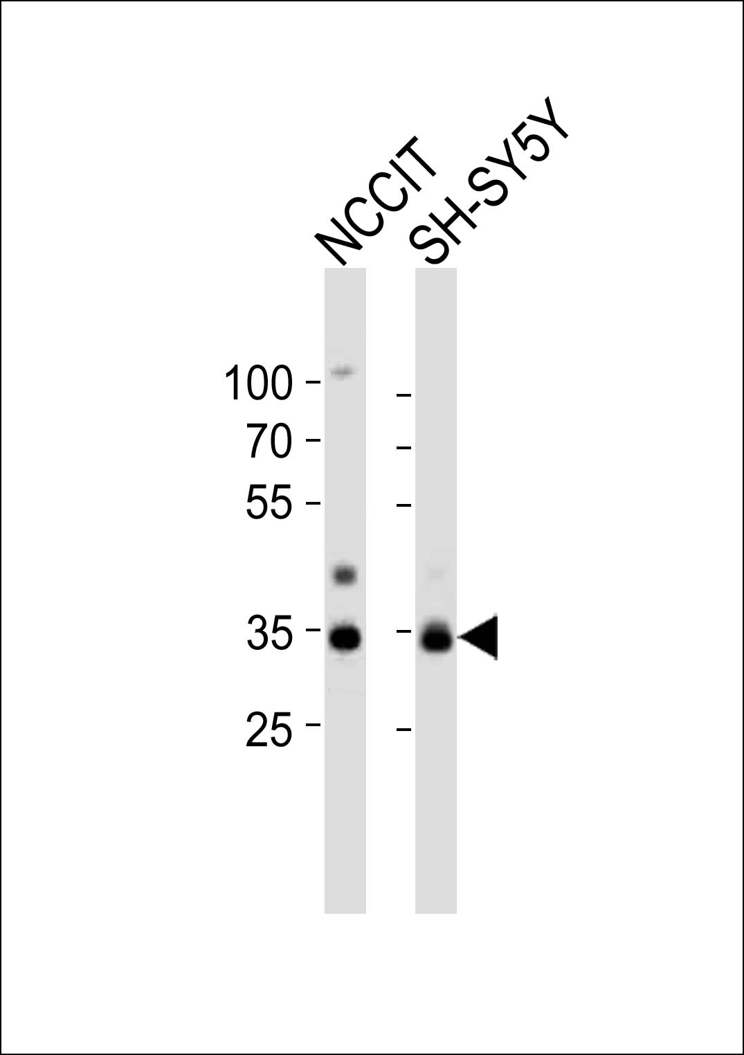 Western blot analysis of lysates from NCCIT, SH-SY5Y cell line (from left to right), using WDR82 Antibody (N-term)(Cat. #AP20978b). AP20978b was diluted at 1:1000 at each lane. A goat anti-rabbit IgG H&L(HRP) at 1:10000 dilution was used as the secondary antibody. Lysates at 20ug per lane.