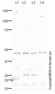
All lanes : Anti-ADFP antibody (ab78920) at 1 µg/mlLane 1 : HeLa (Human epithelial carcinoma cell line) Whole Cell Lysate Lane 2 : Jurkat (Human T cell lymphoblast-like cell line) Whole Cell Lysate Lane 3 : HepG2 (Human hepatocellular liver carcinoma cell line) Whole Cell Lysate Lane 4 : HEK293 (Human embryonic kidney cell line) Whole Cell LysateLysates/proteins at 10 µg per lane.SecondaryGoat polyclonal to Rabbit IgG - H&L - Pre-Adsorbed (HRP) at 1/3000 dilutiondeveloped using the ECL techniquePerformed under reducing conditions.
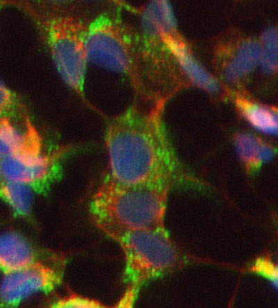
ICC/IF image of ab78920 stained HepG2 cells. The cells were 100% Methanol fixed (5 min) and then incubated in 1%BSA / 10% normal Goat serum / 0.3M glycine in 0.1% PBS-Tween for 1h to permeabilise the cells and block non-specific protein-protein interactions. The cells were then incubated with the antibody (ab78920, 5µg/ml) overnight at +4°C. The secondary antibody (green) was Alexa Fluor® 488 Goat anti-Rabbit IgG (H+L) used at a 1/1000 dilution for 1h. Alexa Fluor® 594 WGA was used to label plasma membranes (red) at a 1/200 dilution for 1h. DAPI was used to stain the cell nuclei (blue) at a concentration of 1.43µM. This antibody also gave a positive result in 100% Methanol fixed (5 min) HeLa, and Hek293 cells at 5µg/ml, and in 4% PFA fixed (10 min) HeLa, Hek293, and HepG2 cells at 1µg/ml
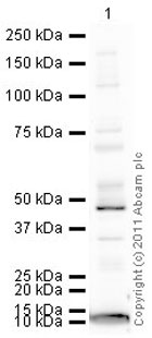
Anti-ADFP antibody (ab78920) at 1 µg/ml + Heart (Mouse) Tissue Lysate at 10 µgSecondaryGoat Anti-Rabbit IgG H&L (HRP) preadsorbed (ab97080) at 1/5000 dilutiondeveloped using the ECL techniquePerformed under reducing conditions.
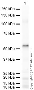
Anti-ADFP antibody (ab78920) at 1 µg/ml + Human ADFP full length protein (ab73631) at 0.1 µgSecondaryGoat Anti-Rabbit IgG H&L (HRP) preadsorbed (ab97080) at 1/5000 dilutiondeveloped using the ECL techniquePerformed under reducing conditions.Exposure time : 1 minute