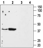
Western blot analysis of mouse kidney (lanes 1 and 3) and mouse heart (lanes 2 and 4) membranes: 1, 2. Anti-Angiotensin II Receptor Type-1 (extracellular) antibody (AG1463), (1:500). 3, 4. Anti-Angiotensin II Receptor Type-1 (extracellular) antibody, preincubated with the control peptide antigen.
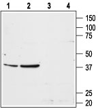
Western blot analysis of rat liver (lanes 1 and 3) and rat kidney (lanes 2 and 4) membranes: 1, 2. Anti-Angiotensin II Receptor Type-1 (extracellular) antibody (AG1463), (1:200). 3, 4. Anti-Angiotensin II Receptor Type-1 (extracellular) antibody, preincubated with the control peptide antigen.
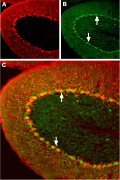
Expression of Angiotensin II Receptor Type-1 in mouse cerebellum Immunohistochemical staining of mouse cerebellum using Anti-Angiotensin II Receptor Type-1 (extracellular) antibody (#AG1463). A. Mouse anti-Parvalbumin (red) is detected in the Purkinje layer. B. In the same section, AT1 receptor (green) is also present in the Purkinje layer. Arrows point at AT1 receptor immunoreactive cells. Merge of A and B pannels reveals partial co-localization.
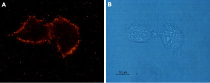
xpression of Angiotensin II Receptor Type-1 in rat C6 glioma cells Immunocytochemical staining of live intact rat C6 glioma cells. A. Cells were stained using Anti-Angiotensin II Receptor Type-1 (extracellular) antibody (#AG1463), (1:100), followed by goat anti-rabbit-AlexaFluor-555 secondary antibody. B. Live intact C6 cells.
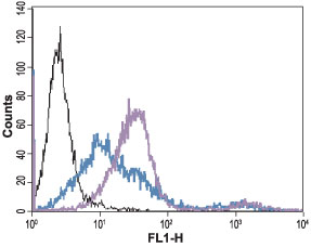
Indirect flow cytometry analysis of live intact human Jurkat T-cell leukemia cells: Black: Unstained cells + goat-anti-rabbit-FITC. Green: Cells + Anti-Angiotensin II Receptor Type-1 (extracellular) antibody (#AG1463), (5 µg) + goat-anti-rabbit-FITC. Green: Cells + Anti-Angiotensin II Receptor Type-1 (extracellular) antibody, (10 µg) + goat-anti-rabbit-FITC.