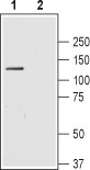
Western blot analysis of rat brain lysate: 1. Anti-NBCe1 (extracellular) antibody (#AG1415), (1:200). 2. Anti-NBCe1 (extracellular) antibody, preincubated with the control peptide antigen.
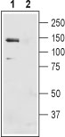
Western blot analysis of mouse liver membranes: 1. Anti-NBCe1 (extracellular) antibody (#AG1415), (1:400). 2. Anti-NBCe1 (extracellular) antibody, preincubated with the control peptide antigen.
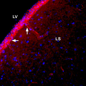
Expression of NBCe1 in rat lateral septum Immunohistochemical staining of immersion-fixed, free floating rat brain frozen sections using Anti-NBCe1 (extracellular) antibody (#AG1415), (1:400). NBCe1 staining (red) is detected in blood vessel outlines (vertical arrow) in the lateral septum (LS) and along the wall of the lateral ventricle (LV) (horizontal arrow). DAPI is used as the counterstain (blue).
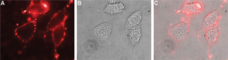
Expression of NBCe1 in rat PC12 cells Immunocytochemical staining of live intact rat PC12 pheochromocytoma cells. A. Extracellular staining of cells with Anti-NBCe1 (extracellular) antibody (#AG1415), (1:100), followed by goat anti-rabbit-AlexaFluor-594 secondary antibody (red). B. Live image of the cells. C. Merge of the two images.