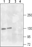
Western blot analysis of rat (lanes 1 and 3) and mouse (lanes 2 and 4) brain lysates: 1, 2. Anti-TrkA (extracellular) antibody (#AG1416), (1:200). 3, 4. Anti-TrkA (extracellular) antibody, preincubated with the control peptide antigen.
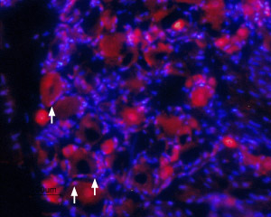
Expression of TrkA in rat DRG Immunohistochemical staining of rat dorsal root ganglia (DRG) frozen sections using Anti-TrkA (extracellular) antibody (#AG1416), (1:100). TrkA (red staining) is expressed in DRG neurons and in satellite microglia (arrows). Hoechst 33342 is used as the counterstain.
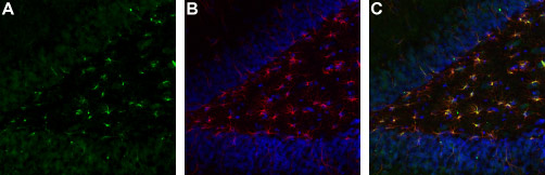
Expression of TrkA in rat brain hippocampal dentate gyrus Immunohistochemical staining of immersion-fixed, free floating rat brain frozen sections. A. Brain sections were stained using Anti-TrkA (extracellular) antibody (#AG1416), (1:1000), (green staining). B. The same section was also stained for glial fibrillary acidic protein (GFAP) (red and counterstained blue). C. Overlay of A and B demonstrates co-localization of TrkA and GFAP in dentate gyrus astrocytes.

Expression of TrkA in live intact rat PC12 cells Immunocytochemical staining of live intact rat PC12 cells. A. Cells were stained with Anti-TrkA (extracellular) antibody (#AG1416) (1:50), followed by goat anti-rabbit-AlexaFluor-494 secondary antibody (red). B. Cell nuclei were visualized with the membrane-permeable DNA dye Hoechst 33342 (blue staining). C. Live view of the cells.
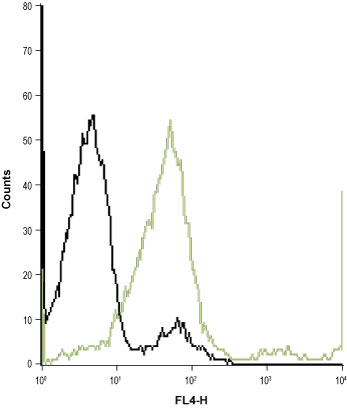
Indirect flow cytometry analysis of live intact Jurkat (human T cell leukemia) cell line: ___ Cells + goat-anti-rabbit-Cy5.___ Cells + Anti-TrkA (extracellular) antibody (#AG1416), (1:25) + goat-anti-rabbit-Cy5.