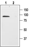
Western blot analysis of rat brain membranes: 1. Anti-Human p75NTR (extracellular) antibody (#AG1417), (1:200). 2. Anti-Human p75NTR (extracellular) antibody, preincubated with the control peptide antigen.
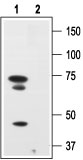
Western blot analysis of human melanoma cells A875: 1. Anti-Human p75NTR (extracellular) antibody (#AG1417), (1:200). 2. Anti-Human p75NTR (extracellular) antibody, preincubated with the control peptide.
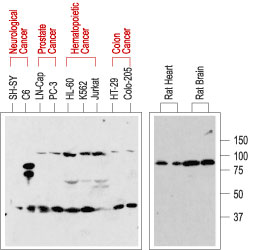
Western blot analysis of normal rat tissue (right) and in human cancer cell lines (left): p75NTR is visualized with Anti-Human p75NTR (extracellular) antibody (#AG1417), (1:200). Note that human cancer cell lines from hemapoietic origin show high p75NTR expression, while cell lines from prostate and colon cancer origin show lower levels. Interestingly, p75NTR from rat (right blot and the C6 cell line) and human (left blot) samples run with a different apparent MW, probably due to species-specific differential glycosylation.
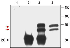
Immunoprecipitation of 3T3/p75NTR transfected cells: 1. Cell lysate + protein A beads. 2. Cell lysate + protein A beads + pre-immune rabbit serum. 3. Cell lysate + protein A beads + Anti-Human p75NTR (extracellular) antibody (#AG1417). 4. Cell lysate. Red arrows indicate the p75NTR receptor while the black arrow shows the IgG heavy chain. Immunoblot was performed with the Anti-Human p75NTR (extracellular) antibody.
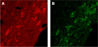
Expression of p75NTR in rat brain Immunohistochemical staining of rat brain with Anti-Human p75NTR (extracellular) antibody (#AG1417). A. Cells in the diagonal band are stained positive for p75NTR. B. Staining of the same section with goat anti-ChAT confirms that p75NTR staining is specific to cholinergic neurons.
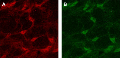
Expression of p75NTR in rat brain Immunohistochemical staining of rat brain with Anti-Human p75NTR (extracellular) antibody (#AG1417). A. Cells in the nucleus basalis mangocellularis are stained positive for p75NTR. B. Staining of the same section with goat anti-ChAT confirms that the p75NTR staining is specific to cholinergic neurons.
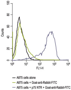
Indirect flow cytometry analysis of A875 human melanoma cells