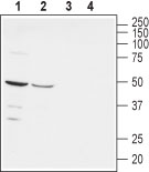
Western blot analysis of rat (lanes 1 and 3) and mouse (lanes 2 and 4) brain lysates: 1-2. Anti-Nectin-1 (extracellular) antibody (#AG1418), (1:200). 3-4. Anti-Nectin-1 (extracellular) antibody, preincubated with the control peptide antigen.

Western blot analysis of human Jurkat T-cell leukemia (lanes 1 and 3) and human MEG-01 chronic myelogenous leukemia (lanes 2 and 4) cell lysates: 1-2. Anti-Nectin-1 (extracellular) antibody (#AG1418), (1:200). 3-4. Anti-Nectin-1 (extracellular) antibody, preincubated with the control peptide antigen.

Expression of Nectin-1 in rat cerebellum Immunohistochemical staining of immersion-fixed, free floating rat brain frozen sections using Anti-Nectin-1 (extracellular) antibody (#AG1418), (1:100). A. Nectin-1 staining (red) is apparent in Purkinje neurons and their dendritic tree (arrow). B. Cell nuclei in the same section are visualized with DAPI (blue). C. Merge of the two images.

Expression of Nectin-1 in rat PC12 cells Immunocytochemical staining of live intact rat PC12 pheochromocytoma cells. A. Extracellular staining of cells with Anti-Nectin-1 (extracellular) antibody (#AG1418), (1:100), followed by goat anti-rabbit-AlexaFluor-594 secondary antibody (red). B. Live image of the cells. C. Merge of the two images.
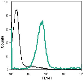
Indirect flow cytometry analysis of live intact human THP-1 monocytic leukemia cell line: ___ Cells + goat anti-rabbit-AlexaFluor-488 secondary antibody.___ Cells + Anti-Nectin-1 (extracellular) antibody (#AG1418), (1:15) + goat anti-rabbit-AlexaFluor-488 secondary antibody.