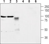
Western blot analysis of rat brain membrane (lanes 1 and 4), mouse brain membrane (lanes 2 and 5) and rat PC-12 cells (lanes 3 and 6): 1-3. Anti-Neuroligin 2 (extracellular) antibody (#AG1419), (1:400). 4-6. Anti-Neuroligin 2 (extracellular) antibody preincubated with the control peptide antigen.
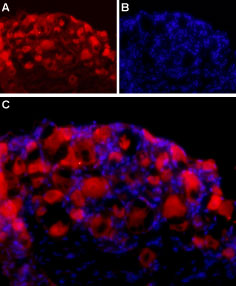
Expression of Neuroligin 2 in rat DRG Immunohistochemical staining of adult rat dorsal root ganglion (DRG) using Anti-Neuroligin 2 (extracellular) antibody (#AG1419) followed by goat anti-rabbit-AlexaFluor-594 secondary antibody. A. Neuroligin 2 labeling (red) appears in the cell bodies of the DRG neurons. B. Nuclear staining using DAPI as the counter stain (blue). C. Merged image of A and B.
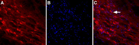
Expression of Neuroligin-2 in rat brain Immunnohistochemical staining of rat reticular thalamic nucleus using Anti-Neuroligin-2 (extracellular) antibody (#AG1419). A. Neuroligin-2 staining (red) is detected in neuronal outlines (arrow). B. Nucleus staining using DAPI as the counterstain. C. Merged images od A and B.
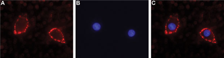
Expression of Neuroligin 2 in rat PC-12 cells Immunocytochemical staining of intact living rat PC-12 cells. A. Extracellular staining of cells using Anti-Neuroligin 2 (extracellular) antibody (#AG1419), (1:25), followed by goat anti-rabbit-AlexaFluor-594 secondary antibody (red). B. Nuclear staining using DAPI as the counterstain (blue). C. Merged images of A and B.