LH-Receptor-extracellular-Antibody
| Name | LH-Receptor-extracellular-Antibody |
|---|---|
| Supplier | Abgent, a WuXi AppTec company |
| Catalog | AG1424 |
| Prices | $375.00, $475.00, $575.00 |
| Sizes | 25 µl, 50 µl, 200 µl |
| Host | Rabbit |
| Clonality | Polyclonal |
| Applications | WB LCI |
| Species Reactivities | Human, Rat |
| Antigen | Lyophilized powder can be stored intact at room temperature for several weeks. For longer periods, it should be stored at -20°C. |
| Purity/Format | Affinity purified antibody, lyophilized powder |
| Description | Rabbit Polyclonal |
| Gene | Lhcgr |
| Supplier Page | Shop |
Product images
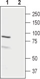 Western blot analysis of rat ovary lysate: 1. Anti-LH Receptor (extracellular) antibody (#AG1424), (1:2000). 2. Anti-LH Receptor (extracellular) antibody, preincubated with the control antigen.
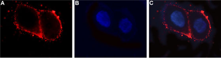 Expression of LH receptor in human ovary cell line Immunocytochemical staining of intact living OVCAR3 cells. A. Extracellular staining of cells with Anti-LH Receptor (extracellular) antibody (#AG1424), (1:25) followed by goat anti-rabbit-AlexaFluor-594 secondary antibody. B. Nuclear staining of cells using DAPI as the counterstain. C. Merged images of A and B.