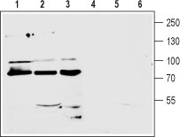
Western blot analysis of mouse (lanes 1 and 4) and rat (lanes 2 and 5) brain membranes and human CCF-STTGI astrocytoma (lanes 3 and 6) cell line lysate (1:200): 1-3. Anti-mGluR7 (extracellular) antibody (#AG1255), (1:200). 4-6. Anti-mGluR7 (extracellular) antibody, preincubated with the control peptide antigen.
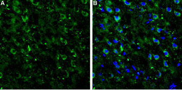
Expression of mGluR7 in rat neocortex Immunohistochemical staining of rat neocortex frozen sections using Anti-mGluR7 (extracellular) antibody (#AG1255). A. mGluR7 staining (green) appears in several neocortical neurons. B. Merge image showing mGluR7 staining together with cell nuclei (blue).
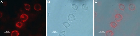
Expression of mGluR7 in rat PC12 cells Immunocytochemical staining of live intact rat pheochromocytoma PC12 cells. A. Cells were stained with Anti-mGluR7 (extracellular) antibody (#AG1255), (1:100), followed by goat anti-rabbit-AlexaFluor-594 secondary antibody (red). B. Live view of the cells. C. Merge of the two pictures.

Indirect flow cytometry analysis of live intact human T cell leukemia (Jurkat) cell line: Black: Unstained cells + goat-anti-rabbit-PE. Green: Cells + Anti-mGluR7 (extracellular) antibody (#AG1255), (1:20) + goat-anti-rabbit-PE.