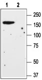
Western blot analysis of rat brain lysate: 1. Anti-mGluR1 (extracellular) antibody (#AG1263), (1:200). 2. Anti-mGluR1 (extracellular) antibody, preincubated with the control peptide antigen.
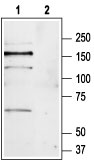
Western blot analysis of mouse brain lysate: 1. Anti-mGluR1 (extracellular) antibody (#AG1263), (1:200). 2. Anti-mGluR1 (extracellular) antibody, preincubated with the control peptide antigen.
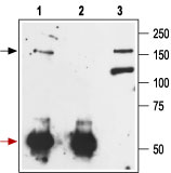
Immunoprecipitation of rat brain lysate: 1. Cell lysate + protein A beads + Anti-mGluR1 (extracellular) antibody (#AG1263). 2. Cell lysate + protein A beads + pre-immune rabbit serum. 3. Cell lysate.
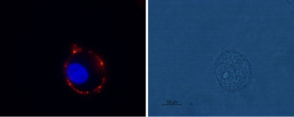
Expression of mGluR1 in rat C6 glioma cells Immunocytochemical staining of live intact rat C6 glioma cells using Anti-mGluR1 (extracellular) antibody (#AG1263), (1:100), followed by goat-anti-rabbit-AlexaFluor-555 secondary antibody (red). Nuclei were stained with Hoechst 33342 (blue).
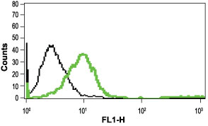
Indirect flow cytometry analysis in live intact Jurkat cells: Black: Unstained cells + goat-anti-rabbit-FITC. Green: Cells + Anti-mGluR1 (extracellular) antibody (#AG1263), (10 µg) + goat-anti-rabbit-FITC.