TRPC1-extracellular-Antibody
| Name | TRPC1-extracellular-Antibody |
|---|---|
| Supplier | Abgent, a WuXi AppTec company |
| Catalog | AG1323 |
| Prices | $375.00, $475.00, $575.00 |
| Sizes | 25 µl, 50 µl, 200 µl |
| Host | Rabbit |
| Clonality | Polyclonal |
| Applications | WB LCI |
| Species Reactivities | Human, Mouse, Rat |
| Antigen | Lyophilized powder can be stored intact at room temperature for several weeks. For longer periods, it should be stored at -20°C. |
| Purity/Format | Affinity purified antibody, lyophilized powder |
| Description | Rabbit Polyclonal |
| Gene | Trpc1 |
| Supplier Page | Shop |
Product images
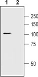 Western blot analysis of rat brain lysate: 1. Anti-TRPC1 (extracellular) antibody (#AG1323), (1:200). 2. Anti-TRPC1 (extracellular) antibody, preincubated with the control peptide antigen.
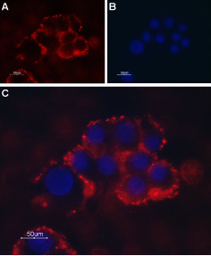 Expression of TRPC1 in PC12 cells Immunocytochemical staining of intact living PC12 cells. A. Extracellular staining of cells with Anti-TRPC1 (extracellular) antibody (#AG1323), (1:50) followed by goat anti-rabbit-AlexaFluor-594 secondary antibody. B. Nuclear staining with DAPI as the counterstain. C. Merged images of A and B.