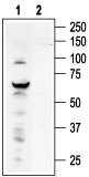
Western blot analysis of ND7/23 cell line lysate: 1. Anti-TRPV4 antibody (#AG1349), (1:200). 2. Anti-TRPV4 antibody, preincubated with the control peptide antigen.
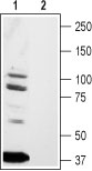
Western blot analysis of rat brain lysates: 1. Anti-TRPV4 antibody (#AG1349), (1:200). 2. Anti-TRPV4 antibody, preincubated with the control peptide antigen.
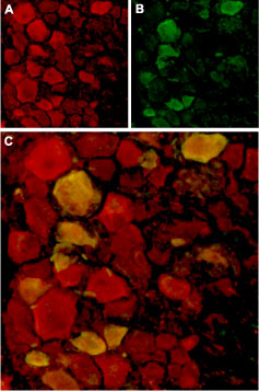
Expression of TRPV4 in rat DRG Immunohistochemical staining of rat dorsal root ganglion (DRG) frozen sections using Anti-TRPV4 antibody (#AG1349). A. TRPV4 (red) in DRG neurons. B. Staining with mouse anti-Parvalbumin (green) in the same DRG section. C. Confocal merge of TRPV4 and Parvalbumin demonstrates colocalization.
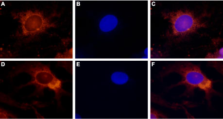
Expression of TRPV4 in rat DRG primary culture Immunocytochemical staining of paraformaldehyde-fixed and permeabilized rat dorsal root ganglion (DRG) primary culture. A, D. Staining using Anti-TRPV4 antibody (#AG1349), (1:500), followed by goat anti-rabbit-AlexaFluor-555 secondary antibody. B, E. Nuclear staining of cells using the cell-permeable dye Hoechst 33342. C. Merged image of panels A and B. F. Merged image of panels D and E.