
Western blot analysis of rat brain membranes: 1. Anti-TRPC4 antibody (#AG1356), (1:200). 2. Anti-TRPC4 antibody, preincubated with the control peptide antigen.
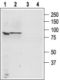
Western blot analysis of PC3 (lanes 1,3) and LNCaP cell lysates: 1. Anti-TRPC4 antibody (#AG1356), (1:200). 2. Anti-TRPC4 antibody, preincubated with the control peptide antigen.
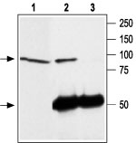
Immunoprecipitation of rat brain lysate: 1. Rat brain lysate. 2. Lysate immunoprecipitated with Anti-TRPC4 antibody (#AG1356), (6 mg). 3. Lysate immunoprecipitated with pre-immune rabbit serum. The upper arrow indicates the TRPC4 channel while the lower arrow indicates the IgG heavy chain. Western blot analysis was performed with Anti-TRPC4 antibody.
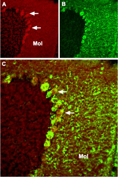
Expression of TRPC4 in mouse cerebellum Immunohistochemical staining of mouse cerebellum frozen sections with Anti-TRPC4 antibody (#AG1356). A. TRPC4 (red) appears in Purkinje cells (arrows) and in the molecular (Mol) layer. B. Staining with mouse anti-parvalbumin (PV) in the same brain section. C. Confocal merge of TRPC4 and PV demonstrates partial co-localization in the Purkinje and the molecular layers.