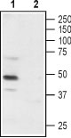
Western blot analysis of rat DRG: 1. Anti-P2X3 Receptor (extracellular) antibody (#AG1060), (1:200). 2. Anti-P2X3 Receptor (extracellular) antibody, preincubated with the control peptide antigen.
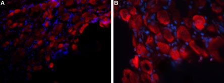
Expression of P2X3 Receptor in rat DRG Immunohistochemical staining of rat dorsal root ganglion (DRG) frozen sections using Anti-P2X3 Receptor (extracellular) antibody (#AG1060), followed by anti-rabbit-AlexaFluor-594 secondary antibody. P2X3 Receptor staining (red) appears in neuronal cell bodies. DAPI was used as the counter stain (blue). A. X20 magnification. B. X40 magnification.
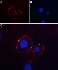
Expression of P2X3 Receptor in rat PC12 cells Immunocytochemical staining of intact living rat pheochromocytoma (PC12) cells. A. Extracellular staining with Anti-P2X3 Receptor (extracellular) antibody (#AG1060), (1:50, red) followed by goat anti-rabbit-AlexaFluor-594 secondary antibody. B. DAPI was used as the counter stain (blue). C. Merge images of A and B.