ab110320 at 1µg/ml staining Aconitase 2 in HDFn cells by Immunocytochemistry (4% paraformaldehyde fixed and 0.1% Triton X-100 permeabilized) followed by Alexa Fluor® 594 goat anti-mouse IgG (H+L) used at a 1/1000 dilution for 1 hour (red). Note: the target protein locates to the mitochondria.
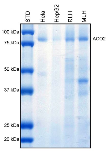
Detection of ab110321 by colloidal Coomassie blue G staining of Immunprecipitate.Staining of Aconitase in HeLa, HepG2, Rat liver homogenate (RLH), and Mouse liver homogenate (MLH) lysates immunoprecipitated using ab110320.
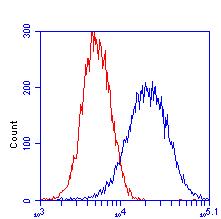
ab110320 at 1µg/ml staining Aconitase 2 in HeLa cells by Flow Cytometry (blue). Isotype control antibody (red).