Anti-Acetylcholinesterase antibody [HR2]
| Name | Anti-Acetylcholinesterase antibody [HR2] |
|---|---|
| Supplier | Abcam |
| Catalog | ab2803 |
| Prices | $388.00 |
| Sizes | 200 µl |
| Host | Mouse |
| Clonality | Monoclonal |
| Isotype | IgG2b |
| Clone | HR2 |
| Applications | ELISA IHC-F IP FC ICC/IF ICC/IF |
| Species Reactivities | Mouse, Rabbit, Guinea Pig, Bovine, Cat, Human, Monkey, Primate |
| Antigen | Full length protein corresponding to Human Acetylcholinesterase |
| Description | Mouse Monoclonal |
| Gene | ACHE |
| Conjugate | Unconjugated |
| Supplier Page | Shop |
Product images
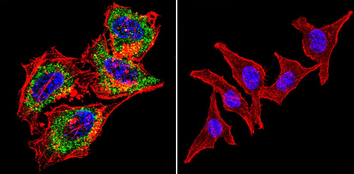 Immunocytochemistry/Immunofluorescence analysis of Acetylcholinesterase shows staining in HeLa cells. Acetylcholinesterase staining (green), F-Actin staining with Phalloidin (red) and nuclei with DAPI (blue) is shown. Cells were grown on chamber slides and fixed with formaldehyde prior to staining. Cells were incubated without (control) or with ab2803 (1:200) overnight at 4°C, washed with PBS and incubated with a DyLight-488 conjugated secondary antibody. Images were taken at 60X magnification.
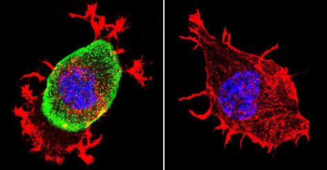 Immunocytochemistry/Immunofluorescence analysis of Acetylcholinesterase shows staining in Neuro-2a cells. Acetylcholinesterase staining (green), F-Actin staining with Phalloidin (red) and nuclei with DAPI (blue) is shown. Cells were grown on chamber slides and fixed with formaldehyde prior to staining. Cells were incubated without (control) or with ab2803 (1:200) overnight at 4°C, washed with PBS and incubated with a DyLight-488 conjugated secondary antibody. Images were taken at 60X magnification.
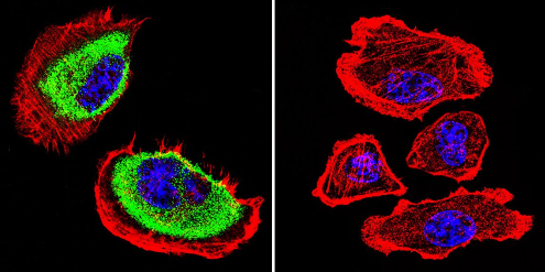 Immunocytochemistry/Immunofluorescence analysis of Acetylcholinesterase shows staining in U251 cells. Acetylcholinesterase staining (green), F-Actin staining with Phalloidin (red) and nuclei with DAPI (blue) is shown. Cells were grown on chamber slides and fixed with formaldehyde prior to staining. Cells were incubated without (control) or with ab2803 (1:200) overnight at 4°C, washed with PBS and incubated with a DyLight-488 conjugated secondary antibody. Images were taken at 60X magnification.
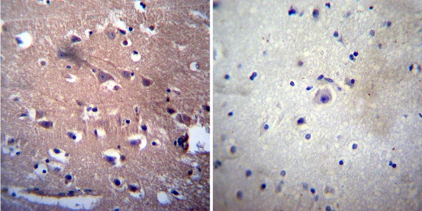 Immunohistochemistry was performed on both normal and cancer biopsies of deparaffinized human Brain tissue. To expose target proteins, heat induced antigen retrieval was performed using 10mM sodium citrate (pH6.0) buffer, microwaved for 8-15 minutes. Following antigen retrieval tissues were blocked in 3% BSA-PBS for 30 minutes at room temperature. Tissues were then probed at a dilution of 1:200 with a mouse monoclonal antibody recognizing Acetylcholinesterase (ab2803) or without primary antibody (negative control) overnight at 4°C in a humidified chamber. Tissues were washed extensively with PBST and endogenous peroxidase activity was quenched with a peroxidase suppressor. Detection was performed using a biotin-conjugated secondary antibody and SA-HRP, followed by colorimetric detection using DAB. Tissues were counterstained with hematoxylin and prepped for mounting.
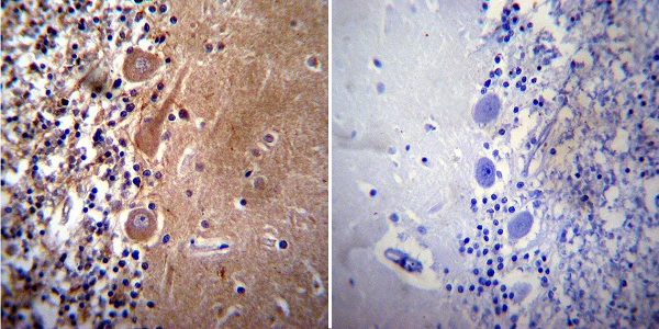 Immunohistochemistry was performed on both normal and cancer biopsies of deparaffinized human Cerebellum tissue. To expose target proteins, heat induced antigen retrieval was performed using 10mM sodium citrate (pH6.0) buffer, microwaved for 8-15 minutes. Following antigen retrieval tissues were blocked in 3% BSA-PBS for 30 minutes at room temperature. Tissues were then probed at a dilution of 1:50 with a mouse monoclonal antibody recognizing Acetylcholinesterase (ab2803) or without primary antibody (negative control) overnight at 4°C in a humidified chamber. Tissues were washed extensively with PBST and endogenous peroxidase activity was quenched with a peroxidase suppressor. Detection was performed using a biotin-conjugated secondary antibody and SA-HRP, followed by colorimetric detection using DAB. Tissues were counterstained with hematoxylin and prepped for mounting.
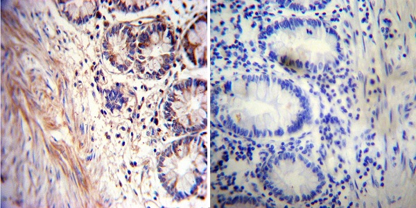 Immunohistochemistry was performed on both normal and cancer biopsies of deparaffinized human Rectum tissue. To expose target proteins, heat induced antigen retrieval was performed using 10mM sodium citrate (pH6.0) buffer, microwaved for 8-15 minutes. Following antigen retrieval tissues were blocked in 3% BSA-PBS for 30 minutes at room temperature. Tissues were then probed at a dilution of 1:20 with a mouse monoclonal antibody recognizing Acetylcholinesterase (ab2803) or without primary antibody (negative control) overnight at 4°C in a humidified chamber. Tissues were washed extensively with PBST and endogenous peroxidase activity was quenched with a peroxidase suppressor. Detection was performed using a biotin-conjugated secondary antibody and SA-HRP, followed by colorimetric detection using DAB. Tissues were counterstained with hematoxylin and prepped for mounting.
![Overlay histogram showing HeLa cells stained with ab2803 (red line). The cells were fixed with 80% methanol (5 min) and then permeabilized with 0.1% PBS-Tween for 20 min. The cells were then incubated in 1x PBS / 10% normal goat serum / 0.3M glycine to block non-specific protein-protein interactions followed by the antibody (ab2803, 1µg/1x106 cells) for 30 min at 22ºC. The secondary antibody used was DyLight® 488 goat anti-mouse IgG (H+L) (ab96879) at 1/500 dilution for 30 min at 22ºC. Isotype control antibody (black line) was mouse IgG2b [PLPV219] (ab91366, 2µg/1x106 cells) used under the same conditions. Unlabelled sample (blue line). Acquisition of >5,000 events were collected using a 20mW Argon ion laser (488nm) and 525/30 bandpass filter. This antibody gave a positive signal in HeLa cells fixed with 4% paraformaldehyde (10 min)/permeabilized with 0.1% PBS-Tween for 20 min used under the same conditions.](http://www.bioprodhub.com/system/product_images/ab_products/2/sub_1/1735_Acetylcholinesterase-Primary-antibodies-ab2803-4.jpg) Overlay histogram showing HeLa cells stained with ab2803 (red line). The cells were fixed with 80% methanol (5 min) and then permeabilized with 0.1% PBS-Tween for 20 min. The cells were then incubated in 1x PBS / 10% normal goat serum / 0.3M glycine to block non-specific protein-protein interactions followed by the antibody (ab2803, 1µg/1x106 cells) for 30 min at 22ºC. The secondary antibody used was DyLight® 488 goat anti-mouse IgG (H+L) (ab96879) at 1/500 dilution for 30 min at 22ºC. Isotype control antibody (black line) was mouse IgG2b [PLPV219] (ab91366, 2µg/1x106 cells) used under the same conditions. Unlabelled sample (blue line). Acquisition of >5,000 events were collected using a 20mW Argon ion laser (488nm) and 525/30 bandpass filter. This antibody gave a positive signal in HeLa cells fixed with 4% paraformaldehyde (10 min)/permeabilized with 0.1% PBS-Tween for 20 min used under the same conditions.
Overlay histogram showing HeLa cells stained with ab2803 (red line). The cells were fixed with 80% methanol (5 min) and then permeabilized with 0.1% PBS-Tween for 20 min. The cells were then incubated in 1x PBS / 10% normal goat serum / 0.3M glycine to block non-specific protein-protein interactions followed by the antibody (ab2803, 1µg/1x106 cells) for 30 min at 22ºC. The secondary antibody used was DyLight® 488 goat anti-mouse IgG (H+L) (ab96879) at 1/500 dilution for 30 min at 22ºC. Isotype control antibody (black line) was mouse IgG2b [PLPV219] (ab91366, 2µg/1x106 cells) used under the same conditions. Unlabelled sample (blue line). Acquisition of >5,000 events were collected using a 20mW Argon ion laser (488nm) and 525/30 bandpass filter. This antibody gave a positive signal in HeLa cells fixed with 4% paraformaldehyde (10 min)/permeabilized with 0.1% PBS-Tween for 20 min used under the same conditions.
Product References
Postmortem changes in the neuroanatomical characteristics of the primate brain: - Postmortem changes in the neuroanatomical characteristics of the primate brain:
Lavenex P, Lavenex PB, Bennett JL, Amaral DG. J Comp Neurol. 2009 Jan 1;512(1):27-51.
Monoclonal antibodies to human brain acetylcholinesterase: properties and - Monoclonal antibodies to human brain acetylcholinesterase: properties and
Rakonczay Z, Brimijoin S. Cell Mol Neurobiol. 1988 Mar;8(1):85-93.