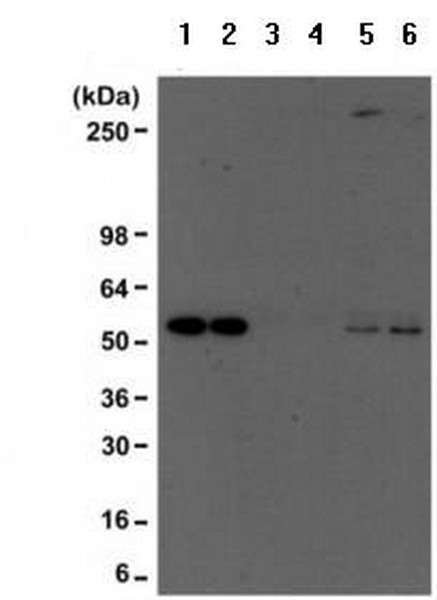
Lane 1: A431 whole cell lysateLane 2: A431 whole cell lysate (pretreated with Trichostatin A)Lane 3: A431 whole cell lysate immunoprecipitated with Rabbit IgGLane 4: A431 whole cell lysate (pretreated with Trichostatin A) immunoprecipitated with Rabbit IgGLane 5: A431 whole cell lysate immunoprecipitated with ab190479 at 1/500Lane 6: A431 whole cell lysate (pretreated with Trichostatin A) immunoprecipitated with ab190479 at 1/500 Western blot performed using anti-PTEN mouse monoclonal antibody.
![All lanes : Anti-Acetylated Lysine antibody [RM101] (ab190479) at 1/2000 dilutionLane 1 : Lysate of nontreated HeLa cellsLane 2 : Lysate of HeLa cells treated with Trichostatin ALane 3 : Lysate of nontreated HeLa cellsLane 4 : Lysate of HeLa cells treated with Trichostatin ALane 5 : Lysate of nontreated HeLa cellsLane 6 : Lysate of HeLa cells treated with Trichostatin Adeveloped using the ECL techniqueExposure time increased from blot on left (lanes 1, 2) to blot on right (lanes 5,6).](http://www.bioprodhub.com/system/product_images/ab_products/2/sub_1/1711_ab190479-225784-ab190479WB1.jpg)
All lanes : Anti-Acetylated Lysine antibody [RM101] (ab190479) at 1/2000 dilutionLane 1 : Lysate of nontreated HeLa cellsLane 2 : Lysate of HeLa cells treated with Trichostatin ALane 3 : Lysate of nontreated HeLa cellsLane 4 : Lysate of HeLa cells treated with Trichostatin ALane 5 : Lysate of nontreated HeLa cellsLane 6 : Lysate of HeLa cells treated with Trichostatin Adeveloped using the ECL techniqueExposure time increased from blot on left (lanes 1, 2) to blot on right (lanes 5,6).
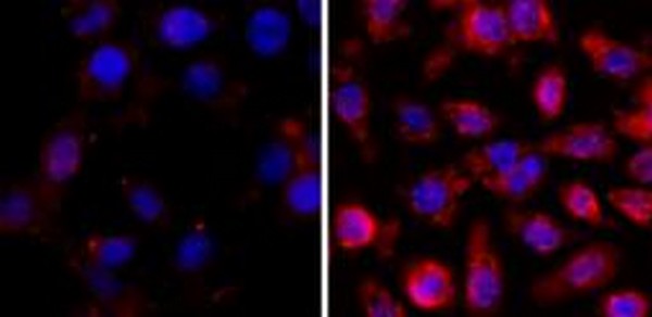
Immunofluorescent analysis of A431cells nontreated (left) or treated with Trichostatin A (right), using ab190479 at 1/500 followed by a PE conjugated secondary antibody (red) and DAPI (blue).