AMT-Antibody-N-term
| Name | AMT-Antibody-N-term |
|---|---|
| Supplier | Abgent, a WuXi AppTec company |
| Catalog | AP6739a |
| Prices | $99.00, $295.00 |
| Sizes | 80 µl, 400 µl |
| Host | Rabbit |
| Clonality | Polyclonal |
| Isotype | Rabbit Ig |
| Applications | WB IHC-P FC ELISA |
| Species Reactivities | Human |
| Antigen | 19-45 aa |
| Purity/Format | Purified polyclonal antibody supplied in PBS with 0.09% (W/V) sodium azide. This antibody is prepared by Saturated Ammonium Sulfate (SAS) precipitation followed by dialysis against PBS. |
| Description | Rabbit Polyclonal |
| Gene | AMT |
| Supplier Page | Shop |
Product images
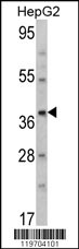 Western blot analysis of AMT Antibody (N-term) (Cat. #AP6739a) in HepG2 cell line lysates (35ug/lane). AMT (arrow) was detected using the purified Pab.
 Formalin-fixed and paraffin-embedded human hepatocarcinoma reacted with AMT Antibody (N-term), which was peroxidase-conjugated to the secondary antibody, followed by DAB staining. This data demonstrates the use of this antibody for immunohistochemistry; clinical relevance has not been evaluated.
 AMT Antibody (N-term) (Cat.#AP6739a) flow cytometry analysis of HepG2 cells (bottom histogram) compared to a negative control cell (top histogram). FITC-conjugated goat-anti-rabbit secondary antibodies were used for the analysis.