![All lanes : Anti-ABCE1 antibody [EPR15373(B)] - C-terminal (ab185548) at 1/5000 dilutionLane 1 : 293 cell lysateLane 2 : K562 cell lysateLane 3 : HeLa cell lysateLysates/proteins at 20 µg per lane.SecondaryGoat Anti-Rabbit IgG, (H+L), Peroxidase conjugated at 1/1000 dilution](http://www.bioprodhub.com/system/product_images/ab_products/2/sub_1/1108_ab185548-219631-ab1855481.jpg)
All lanes : Anti-ABCE1 antibody [EPR15373(B)] - C-terminal (ab185548) at 1/5000 dilutionLane 1 : 293 cell lysateLane 2 : K562 cell lysateLane 3 : HeLa cell lysateLysates/proteins at 20 µg per lane.SecondaryGoat Anti-Rabbit IgG, (H+L), Peroxidase conjugated at 1/1000 dilution
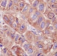
Immunohistochemical analysis of paraffin embedded Human breast carcinoma tissue sections labeling ABCE1 using ab185548 at a 1/100 dilution. A ready to use HRP Polymer for Rabbit IgG was used as the secondary. Hematoxylin counterstain.
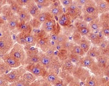
Immunohistochemical analysis of paraffin embedded mouse liver tissue sections labeling ABCE1 using ab185548 at a 1/100 dilution. A ready to use HRP Polymer for Rabbit IgG was used as the secondary. Hematoxylin counterstain.
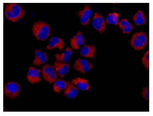
Immunofluorescent analysis of formaldehyde fixed HeLa cells labeling ABCE1 using ab185548 at a 1/250 dilution. A Goat anti rabbit IgG (Alexa Fluor®555) was used as the secondary at a 1/200 dilution.
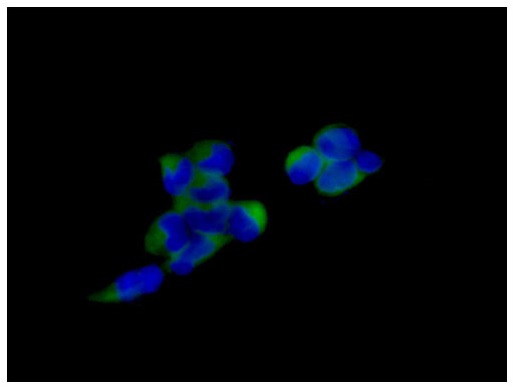
Immunofluorescent analysis of formaldehyde fixed 293 cells labeling ABCE1 using ab185548 at a 1/100 dilution. A Goat anti rabbit IgG (Alexa Fluor®488) was used as the secondary at a 1/200 dilution.
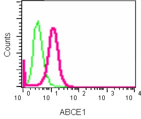
Flow Cytometry analysis of K562 cells labeling ABCE1 using ab185548 at a 1/220 dilution (pink). Goat anti rabbit IgG (FITC) used as the secondary at a 1/150 dilution. Isotype control Rabbit monoclonal IgG (green). Cells were fixed in 2% paraformaldehyde.
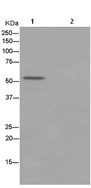
Western blot analysis of ABCE1 in immunoprecipitation pellets from 293 lysate (Lane 1) or 1XPBS (negative control) (Lane 2) using ab185548 at 1/50 dilution. Secondary antibody, Anti-Rabbit IgG (HRP), specific to the non-reduced form of IgG at a 1/1500 dilution.