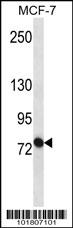
ABCB10 Antibody (S16) (Cat. #AP6109a) western blot analysis in MCF-7 cell line lysates (35ug/lane).This demonstrates the ABCB10 antibody detected the ABCB10 protein (arrow).

ABCB10 antibody (N-term) (Cat. #AP6109a)immunohistochemistry analysis in formalin fixed and paraffin embedded human liver tissue followed by peroxidase conjugation of the secondary antibody and DAB staining.This data demonstrates the use of ABCB10 antibody (N-term) for immunohistochemistry. Clinical relevance has not been evaluated.

All lanes : Anti-ABCB10 Antibody (S16) at 1:1000-1:2000 dilution Lane 1: K562 whole cell lysates Lane 2: Raji whole cell lysates Lysates/proteins at 20 µg per lane. Secondary Goat Anti-Rabbit IgG, (H+L), Peroxidase conjugated at 1/10000 dilution Predicted band size : 79 kDa Blocking/Dilution buffer: 5% NFDM/TBST.

AP6109a staining hABCB10 in Human skeletal muscle tissue sections by Immunohistochemistry (IHC-P - paraformaldehyde-fixed, paraffin-embedded sections). Tissue was fixed with formaldehyde and blocked with 3% BSA for 0. 5 hour at room temperature; antigen retrieval was by heat mediation with a citrate buffer (pH6). Samples were incubated with primary antibody (1/25) for 1 hours at 37°C. A undiluted biotinylated goat polyvalent antibody was used as the secondary antibody.