β-Actin (8H10D10) Mouse mAb
| Name | β-Actin (8H10D10) Mouse mAb |
|---|---|
| Supplier | Cell Signaling Technology |
| Catalog | 3700 |
| Prices | $99.00, $246.00 |
| Sizes | 20 µl (2 western blots), 100 µl (10 western blots) |
| Host | Mouse |
| Clonality | Monoclonal |
| Isotype | IgG2b |
| Clone | 8H10D10 |
| Applications | WB IHC-P ICC/IF FC |
| Species Reactivities | Human, Mouse, Rat, Hamster, Monkey, Dog |
| Antigen | Monoclonal antibody is produced by immunizing animals with a synthetic peptide corresponding to amino-terminal residues of human β-actin. |
| Description | Mouse Monoclonal |
| Gene | ACTB |
| Supplier Page | Shop |
Product images
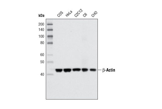 Western blot analysis of extracts from various cell types using β-Actin (8H10D10) Mouse mAb.
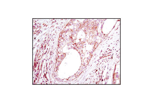 Immunohistochemical analysis of paraffin-embedded human breast carcinoma using β-Actin (8H10D10) Mouse mAb.
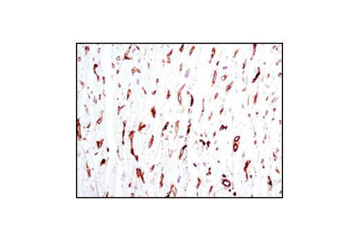 Immunohistochemical analysis of paraffin-embedded human heart using β-Actin (8H10D10) Mouse mAb. Note the lack of staining of cardiac muscle.
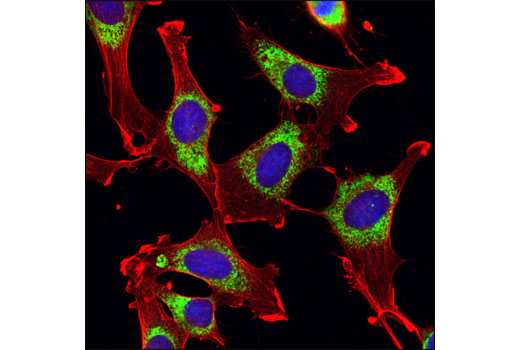 Confocal immunofluorescent analysis of NIH/3T3 cells using β-Actin (8H10D10) Mouse mAb (red) and PDI (C81H6) Rabbit mAb #3501 (green). Blue pseudocolor = DRAQ5 ® #4084 (fluorescent DNA dye).
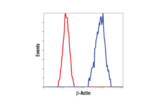 Flow cytometric analysis of HeLa cells using β-Actin (8H10D10) Mouse mAb (blue) compared to a nonspecific negative control antibody (red).