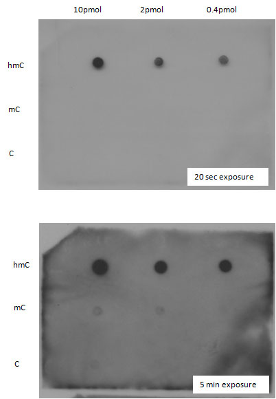
Dot blot assay shows that ab106918 specifically recognized 5-hydroxymethyl Cytidine (hmC). Indicated amounts of hmC, methyl Cytidine (mC) and Cytidine (C) were spotted onto a membrane that was then incubated with ab106918. hmC, mC and C were generated in the following way: M13mp18 DNA had been amplified using primers F and R; F: atttccatgagcgtttttcc R: gcaaggcaaagaattagcaa. A 200 uM dNTP end concentration was used with 1. A,G,C,T and 2. A,G,hmC,T; where C had been replaced with HmdCTP. DNA was in vitro methylated with SssI and SAM, and 2ul of pmol of each base was denatured at 95C for 5 min and spotted and dried onto the membrane. The dot blot membrane was blocked with 10%skimmed milk + 1%BSA blocking overnight and then incubated with ab106918 at 1:500 in blocking solution. A goat anti rat HRP secondary antibody was used for ECL detection. This image is from an anonymous collaborator.
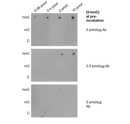
Dot blot competition assay in which ab106918 was preincubated with 5-hydroxymethyl Cytidine (5hmC) at amounts indicated in figure. Specified amounts of 5hmC, methyl Cytidine (mC) and Cytidine (C) were spotted onto membranes and were then incubated with ab106918 that had been preincubated with 5hmC as shown in figure. ab106918 specifically recognized 5hmC and this was blocked by preincubation with 5hmC at 5 pmol/ug ab106918 (Ab). This image is from an anonymous collaborator.
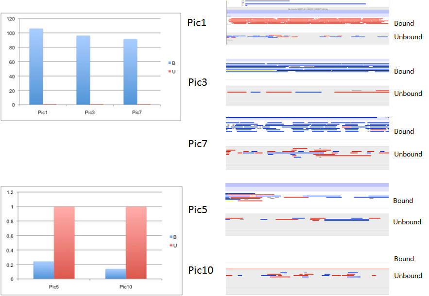
The specificity of ab106918 was confirmed by (h)MeDIP using qPCR validation of regions in ES cells that are highly enriched in 5-hydroxymethyl Cytidine (5hmC) (Pic1, Pic3 and Pic7) or not (Pic5 and Pic10). This image is from an anonymous collaborator.
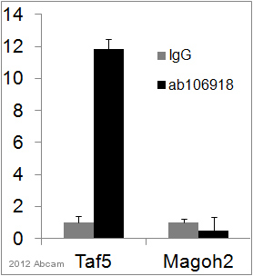
ChIP analysis of mouse ES nuclear cell lysate using ab106918 to bind 5-hydroxymethyl Cytidine. Chromatin was obtained by incubating with primary antibody (0.5 µg/µg chromatin in a glycerol IP buffer) for 16 hours at 4°C. Protein binding was detected using real-time PCR. See Abreview