VILIP3-Antibody-N-term
| Name | VILIP3-Antibody-N-term |
|---|---|
| Supplier | Abgent, a WuXi AppTec company |
| Catalog | AP1563a |
| Prices | $99.00, $295.00 |
| Sizes | 80 µl, 400 µl |
| Host | Rabbit |
| Clonality | Polyclonal |
| Isotype | Rabbit Ig |
| Applications | WB IHC-P ELISA |
| Species Reactivities | Human, Mouse |
| Antigen | 2-32 aa |
| Purity/Format | Purified polyclonal antibody supplied in PBS with 0.09% (W/V) sodium azide. This antibody is prepared by Saturated Ammonium Sulfate (SAS) precipitation followed by dialysis against PBS. |
| Description | Rabbit Polyclonal |
| Gene | HPCAL1 |
| Supplier Page | Shop |
Product images
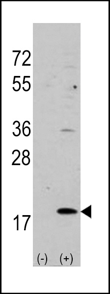 Western blot analysis of VILIP3 (arrow) using rabbit polyclonal VILIP3 Antibody (N-term) (Cat.#AP1563a). 293 cell lysates (2 ug/lane) either nontransfected (Lane 1) or transiently transfected with the VILIP3 gene (Lane 2) (Origene Technologies).
 The anti-VILIP3 Pab (Cat. #AP1563a) is used in Western blot to detect VILIP3 in mouse cerebellum tissue lysate.
 Formalin-fixed and paraffin-embedded human cancer tissue reacted with the primary antibody, which was peroxidase-conjugated to the secondary antibody, followed by AEC staining. This data demonstrates the use of this antibody for immunohistochemistry; clinical relevance has not been evaluated. BC = breast carcinoma