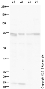
All lanes : Anti-15 Lipoxygenase 1 antibody (ab150051) at 1 µg/mlLane 1 : Kidney (Mouse) Tissue Lysate Lane 2 : Heart (Mouse) Tissue LysateLane 3 : Small Intestine (Mouse) Tissue LysateLane 4 : RAW 264.7 (Mouse leukaemic monocyte macrophage cell line) Whole Cell LysateLysates/proteins at 10 µg per lane.SecondaryGoat Anti-Rabbit IgG H&L (HRP) preadsorbed (ab97080) at 1/5000 dilutiondeveloped using the ECL techniquePerformed under reducing conditions.
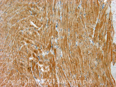
IHC image of 15 Lipoxygenase staining in Mouse normal heart formalin fixed paraffin embedded tissue section, performed on a Leica BondTM system using the standard protocol B. The section was pre-treated using heat mediated antigen retrieval with EDTA buffer (epitope retrieval solution 2) for 20 mins. The section was then incubated with ab150051, 10µg/ml, for 15 mins at room temperature. A Goat anti-Rabbit biotinylated secondary antibody was used to detect the primary, and visualized using an HRP conjugated ABC system. DAB was used as the chromogen. The section was then counterstained with haematoxylin and mounted with DPX. For other IHC staining systems (automated and non-automated) customers should optimize variable parameters such as antigen retrieval conditions, primary antibody concentration and antibody incubation times.