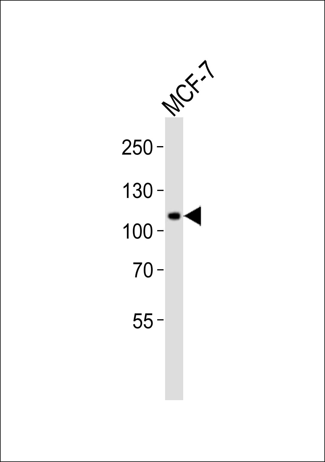
Western blot analysis of lysate from MCF-7 cell line, using RHBDF2 Antibody (N-term)(Cat. #AP13588a). AP13588a was diluted at 1:1000. A goat anti-rabbit IgG H&L(HRP) at 1:10000 dilution was used as the secondary antibody. Lysate at 20ug.
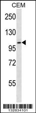
RHBDF2 Antibody (N-term) (Cat. #AP13588a) western blot analysis in CEM cell line lysates (35ug/lane).This demonstrates the RHBDF2 antibody detected the RHBDF2 protein (arrow).
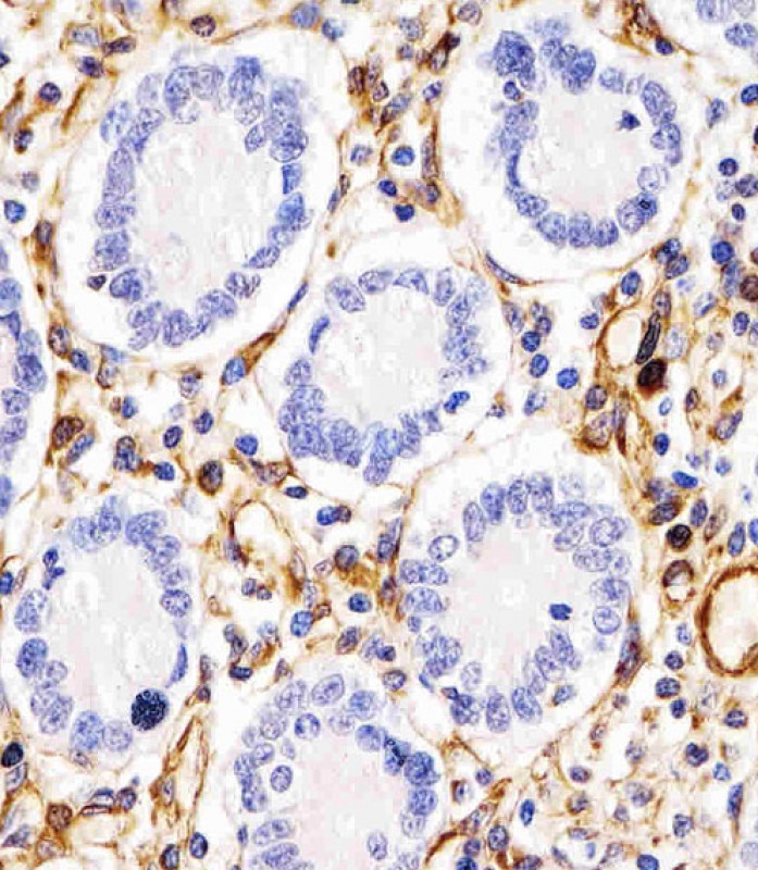
AP13588a staining RHBDF2 in human duodenum tissue sections by Immunohistochemistry (IHC-P - paraformaldehyde-fixed, paraffin-embedded sections). Tissue was fixed with formaldehyde and blocked with 3% BSA for 0. 5 hour at room temperature; antigen retrieval was by heat mediation with a citrate buffer (pH6). Samples were incubated with primary antibody (1/25) for 1 hours at 37°C. A undiluted biotinylated goat polyvalent antibody was used as the secondary antibody.
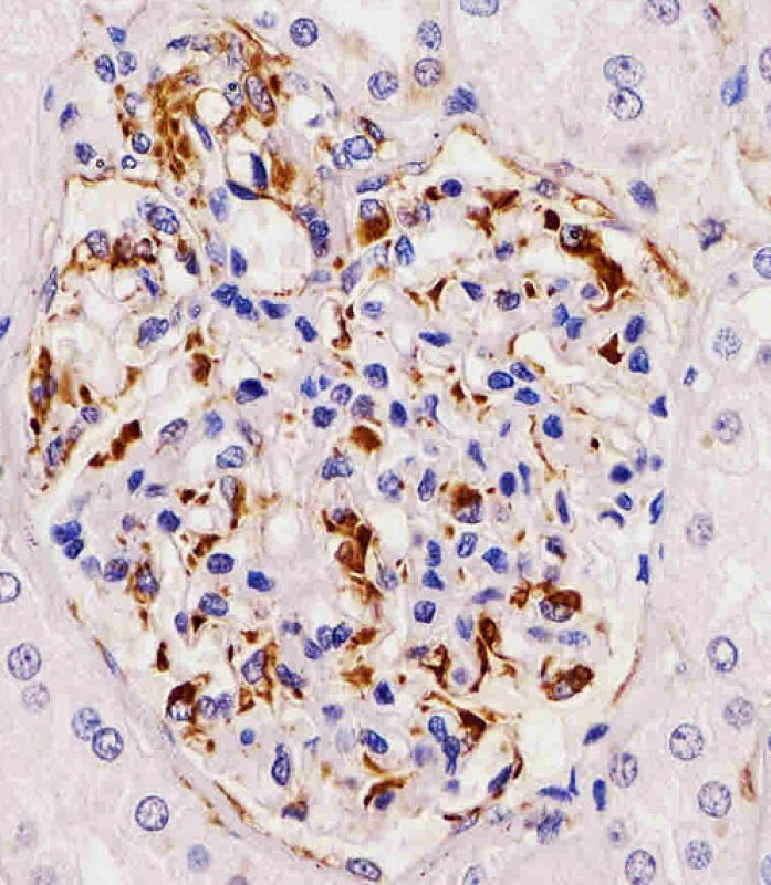
AP13588a staining RHBDF2 in human kidney tissue sections by Immunohistochemistry (IHC-P - paraformaldehyde-fixed, paraffin-embedded sections). Tissue was fixed with formaldehyde and blocked with 3% BSA for 0. 5 hour at room temperature; antigen retrieval was by heat mediation with a citrate buffer (pH6). Samples were incubated with primary antibody (1/25) for 1 hours at 37°C. A undiluted biotinylated goat polyvalent antibody was used as the secondary antibody.
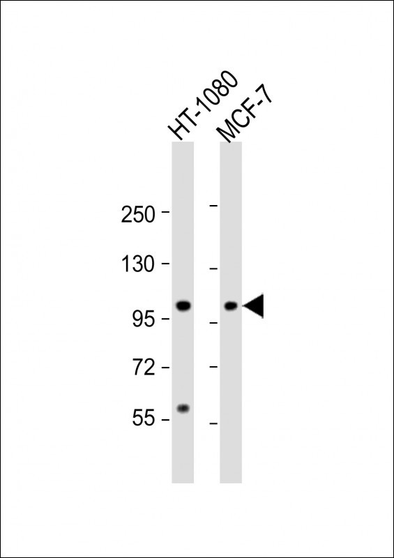
All lanes : Anti-RHBDF2 Antibody (N-term) at 1:2000 dilution Lane 1: HT-1080 whole cell lysate Lane 2: MCF-7 whole cell lysate Lysates/proteins at 20 µg per lane. Secondary Goat Anti-Rabbit IgG, (H+L), Peroxidase conjugated at 1/10000 dilution. Predicted band size : 97 kDa Blocking/Dilution buffer: 5% NFDM/TBST.
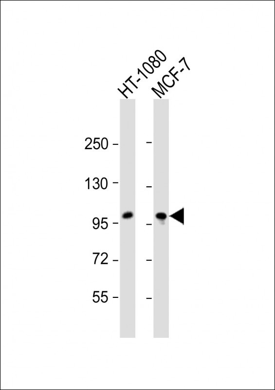
All lanes : Anti-RHBDF2 Antibody (N-term) at 1:2000 dilution Lane 1: HT-1080 whole cell lysate Lane 2: MCF-7 whole cell lysate Lysates/proteins at 20 µg per lane. Secondary Goat Anti-Rabbit IgG, (H+L), Peroxidase conjugated at 1/10000 dilution. Predicted band size : 97 kDa Blocking/Dilution buffer: 5% NFDM/TBST.