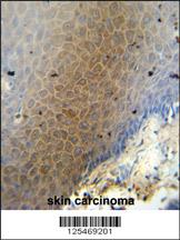
CEP70 antibody (Center) (Cat. #AP10646c) immunohistochemistry analysis in formalin fixed and paraffin embedded human skin carcinoma followed by peroxidase conjugation of the secondary antibody and DAB staining. This data demonstrates the use of the CEP70 antibody (Center) for immunohistochemistry. Clinical relevance has not been evaluated.
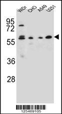
CEP70 Antibody (Center) (Cat. #AP10646c) western blot analysis in WiDr,CHO,A549,U251 cell line lysates (35ug/lane).This demonstrates the CEP70 antibody detected the CEP70 protein (arrow).
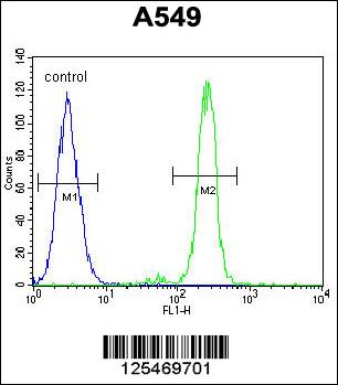
CEP70 Antibody (Center) (Cat. #AP10646c) flow cytometric analysis of A549 cells (right histogram) compared to a negative control cell (left histogram).FITC-conjugated goat-anti-rabbit secondary antibodies were used for the analysis.