CPLX3-Antibody-Center
| Name | CPLX3-Antibody-Center |
|---|---|
| Supplier | Abgent, a WuXi AppTec company |
| Catalog | AP10652c |
| Prices | $99.00, $295.00 |
| Sizes | 80 µl, 400 µl |
| Host | Rabbit |
| Clonality | Polyclonal |
| Isotype | Rabbit Ig |
| Applications | WB IHC-P ELISA |
| Species Reactivities | Human |
| Antigen | 52-81 aa |
| Purity/Format | Purified polyclonal antibody supplied in PBS with 0.09% (W/V) sodium azide. This antibody is purified through a protein A column, followed by peptide affinity purification. |
| Description | Rabbit Polyclonal |
| Gene | CPLX3 |
| Supplier Page | Shop |
Product images
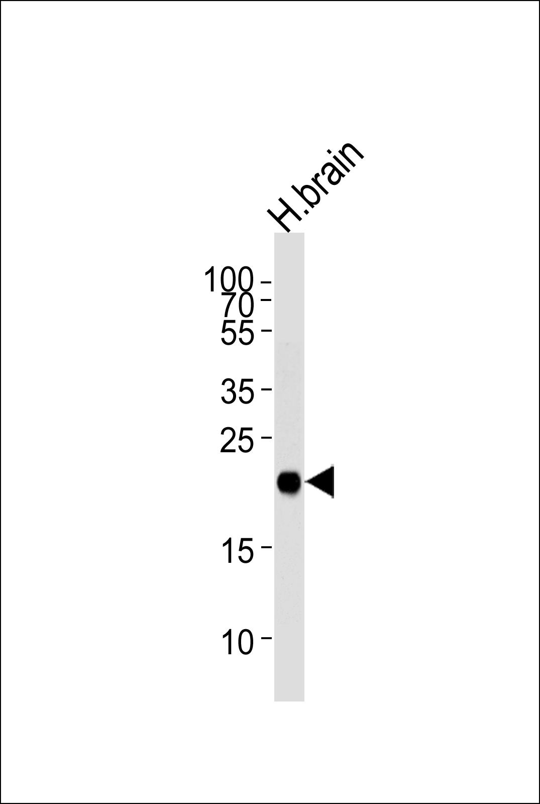 Western blot analysis of lysate from human brain tissue lysate, using CPLX3 Antibody (Center)(Cat. #AP10652c). AP10652c was diluted at 1:1000 at each lane. A goat anti-rabbit IgG H&L(HRP) at 1:5000 dilution was used as the secondary antibody. Lysate at 35ug per lane.
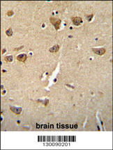 CPLX3 Antibody (Center) (Cat. #AP10652c) immunohistochemistry analysis in formalin fixed and paraffin embedded human brain tissue followed by peroxidase conjugation of the secondary antibody and DAB staining. This data demonstrates the use of the CPLX3 Antibody (Center) for immunohistochemistry. Clinical relevance has not been evaluated.