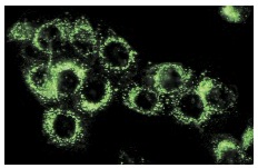
β-dystroglycan (4F7): sc-33702. Immunofluorescence staining of methanol-fixed L6 cells showing membrane localization.
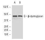
β-dystroglycan (4F7): sc-33702. Western blot analysis of β-dystroglycan expression in C6 (A) and L6 (B) whole cell lysates.
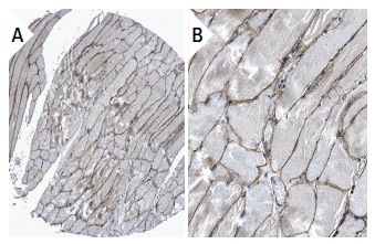
β-dystroglycan (4F7): sc-33702. Immunoperoxidase staining of formalin fixed, paraffin-embedded human skeletal muscle tissue showing membrane staining of myocytes (low and high magnification). Kindly provided by The Swedish Human Protein Atlas (HPA) program.
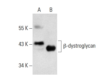
β-dystroglycan (4F7): sc-33702. Western blot analysis of β-dystroglycan expression in rat skeletal muscle tissue extract (A) and SK-BR-3 whole cell lysate (B).