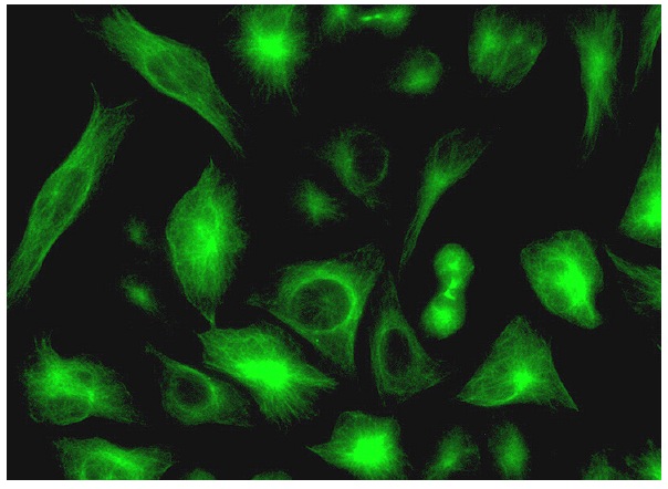
α Tubulin (B-7): sc-5286. Immunofluorescence staining of formalin-fixed HeLa cells showing cytoskeletal localization. Kindly provided by Yang Xiang, Ph.D., Division of Newborn Medicine, Boston Childrens Hospital, Cell Biology Department, Harvard Medical School.
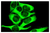
αTubulin (B-7): sc-5286. Immunofluorescence staining of methanol-fixed HeLa cells showing cytoplasmic staining.
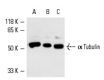
α Tubulin (B-7): sc-5286. Western blot analysis of α Tubulin expression in HeLa (A), K-562 (B) and PC-12 (C) whole cell lysates.
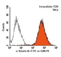
α Tubulin (B-7) PE: sc-5286 PE. Intracellular FCM analysis of fixed and permeabilized HeLa cells. Black line histogram represents the isotype control, normal mouse IgG
2a: sc-2867.
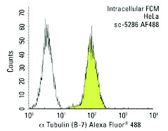
α Tubulin (B-7) Alexa Fluor 488: sc-5286 AF488. Intracellular FCM analysis of fixed and permeabilized HeLa cells. Black line histogram represents the isotype control, normal mouse IgG
2a: sc-3891.
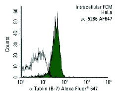
α Tubulin (B-7) Alexa Fluor 647: sc-5286 AF647. Intracellular FCM analysis of fixed and permeabilized HeLa cells. Black line histogram represents the isotype control, normal mouse IgG
2a: sc-24637.
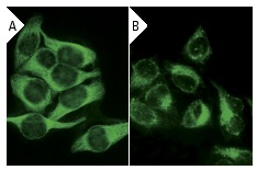
α Tubulin (B-7): sc-5286. Immunofluorescence staining of methanol-fixed HeLa cells showing cytoplasmic localization using indirect FITC (A) staining and direct Alexa Fluor 488 (B) staining.
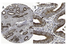
α Tubulin (B-7): sc-5286. Immunoperoxidase staining of formalin fixed, paraffin-embedded human endometrium tissue showing cytoplasmic staining of glandular cells at low (A) and high (B) magnification. Kindly provided by The Swedish Human Protein Atlas (HPA) program.
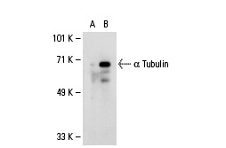
α Tubulin (B-7): sc-5286. Western blot analysis of α Tubulin expression in non-transfected: sc-110760 (A) and human α Tubulin transfected: sc-110978 (B) 293 whole cell lysates.
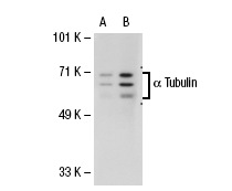
α Tubulin (B-7): sc-5286. Western blot analysis of α Tubulin expression in non-transfected: sc-110760 (A) and human α Tubulin transfected: sc-112325 (B) 293 whole cell lysates.
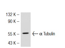
α Tubulin (B-7): sc-5286. Western blot analysis of α Tubulin expression in HeLa whole cell lysate.
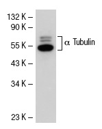
α Tubulin (B-7): sc-5286. Western blot analysis of α Tubulin expression in HeLa whole cell lysate.
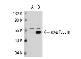
α Tubulin (B-7): sc-5286. Western blot analysis of α4a Tubulin expression in non-transfected: sc-117752 (A) and mouse α4a Tubulin transfected: sc-118128 (B) 293T whole cell lysates.
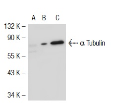
α Tubulin (B-7): sc-5286. Western blot analysis of α8 Tubulin expression in non-transfected 293T: sc-117752 (A), mouse α8 Tubulin transfected 293T: sc-118133 (B) and NIH/3T3 (C) whole cell lysates.
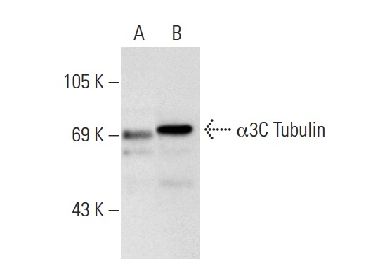
α Tubulin (B-7): sc-5286. Western blot analysis of α3C Tubulin expression in non-transfected: sc-117752 (A) and human α3C Tubulin transfected: sc-173307 (B) 293T whole cell lysates.
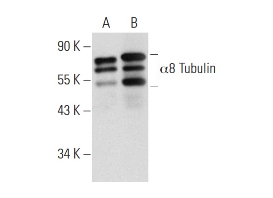
α Tubulin (B-7): sc-5286. Western blot analysis of α8 Tubulin expression in non-transfected: sc-117752 (A) and human α8 Tubulin transfected: sc-127878 (B) 293T whole cell lysates.
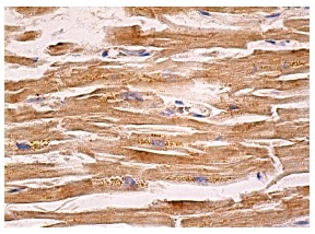
α Tubulin (B-7): sc-5286. Immunoperoxidase staining of formalin fixed, paraffin-embedded human heart muscle tissue showing cytoplasmic staining of myocytes.
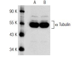
Western blot analysis of α Tubulin expression in untreated (A) and Oxamflatin (sc-205960) treated (B) HeLa whole cell lysates. Antibodies tested include α Tubulin (B-7): sc-5286 (A,B).
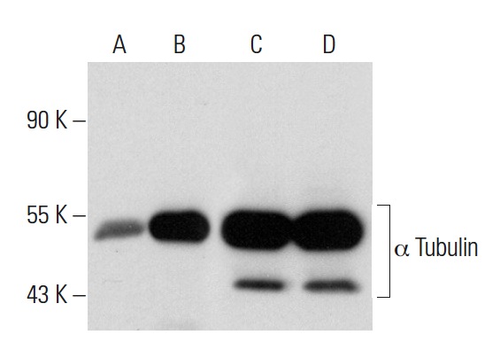
Western blot analysis of α Tubulin expression in untreated (A,C) and Panobinostat (sc-208148) treated (B,D) A549 whole cell lysates. Antibodies tested include acetylated α Tubulin (6-11B-1): sc-23950 (A,B) and α Tubulin (B-7): sc-5286 (C,D).
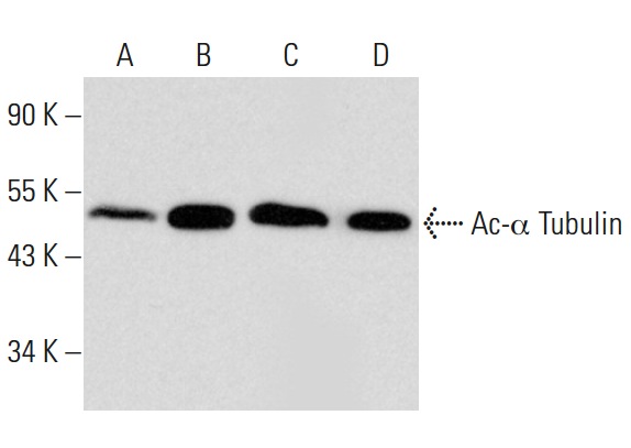
Western blot analysis of α Tubulin acetylation in untreated (A) and CBHA (sc-205240) treated (B) A549 whole cell lysates. Antibodies tested include acetylated α Tubulin (6-11B-1): sc-23950 (A,B) and α Tubulin (B-7): sc-5286 (C,D).
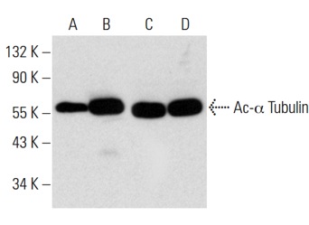
Western blot analysis of α Tubulin acetylation in untreated (A,C) and Scriptaid (sc-202807) treated (B,D) A549 whole cell lysates. Antibodies tested include acetylated α Tubulin (6-11B-1): sc-23950 (A,B) and α Tubulin (B-7): sc-5286 (C,D).


