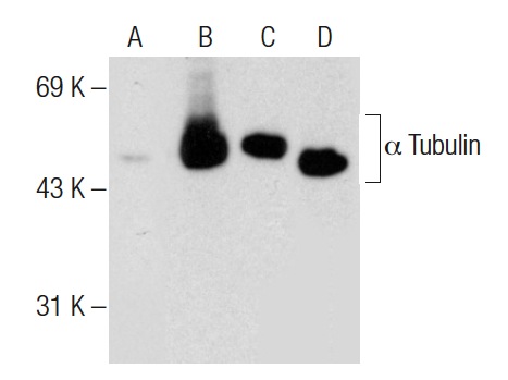
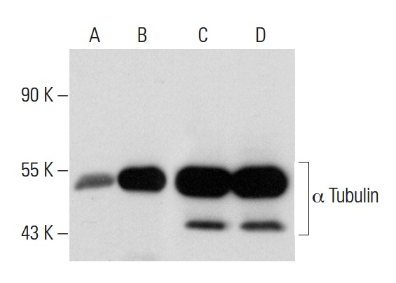
Western blot analysis of α Tubulin acetylation in untreated (A,C) and deacetylase inhibitor cocktail (sc-362323) treated (B,D) NIH/3T3 whole cell lysates. Antibodies tested include acetylated α Tubulin (6-11B-1): sc-23950 (A,B) and α Tubulin (DM1A): sc-32293 (C,D).
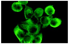
acetylated α Tubulin (6-11B-1): sc-23950. Immunofluorescence staining of methanol-fixed HeLa cells showing cytoplasmic localization.
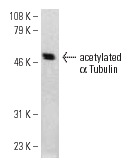
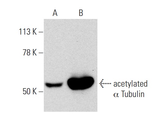
acetylated α Tubulin (6-11B-1): sc-23950. Western blot analysis of acetylated α Tubulin expression in NIH/3T3 whole cell lysate.
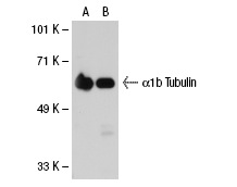
acetylated α Tubulin (6-11B-1): sc-23950. Western blot analysis of α1b Tubulin expression in non-transfected: sc-117752 (A) and human α1b Tubulin transfected: sc-113442 (B) 293T whole cell lysates.
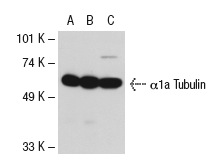
acetylated α Tubulin (6-11B-1): sc-23950. Western blot analysis of α1a Tubulin expression in non-transfected 293T: sc-117752 (A), human α1a Tubulin transfected 293T: sc-116290 (B) and HeLa (C) whole cell lysates.
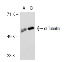
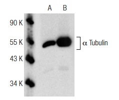
acetylated α Tubulin (6-11B-1): sc-23950. Western blot analysis of α Tubulin expression in non-transfected: sc-117752 (A) and mouse α Tubulin transfected: sc-118096 (B) 293T whole cell lysates.

acetylated α Tubulin (6-11B-1): sc-23950. Western blot analysis of α8 Tubulin expression in non-transfected 293T: sc-117752 (A), mouse α8 Tubulin transfected 293T: sc-118133 (B) and NIH/3T3 (C) whole cell lysates.
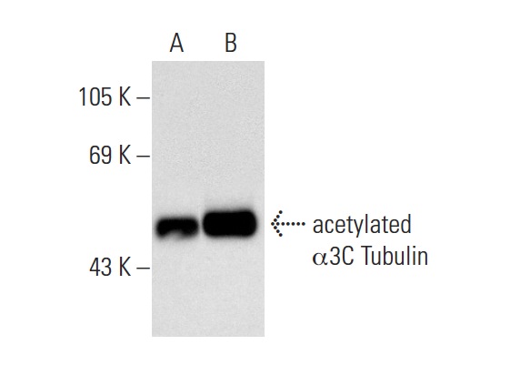
acetylated α Tubulin (6-11B-1): sc-23950. Western blot analysis of α3C Tubulin expression in non-transfected: sc-117752 (A) and human α3C Tubulin transfected: sc-173307 (B) 293T whole cell lysates.
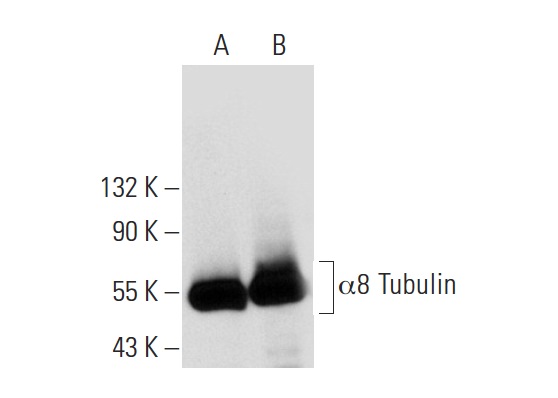
acetylated α Tubulin (6-11B-1): sc-23950. Western blot analysis of α8 Tubulin expression in non-transfected: sc-117752 (A) and human α8 Tubulin transfected: sc-127878 (B) 293T whole cell lysates.
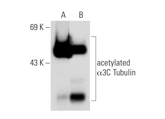
acetylated α Tubulin (6-11B-1): sc-23950. Western blot analysis of α3C Tubulin expression in non-transfected: sc-117752 (A) and human α3C Tubulin transfected: sc-173019 (B) 293T whole cell lysates.
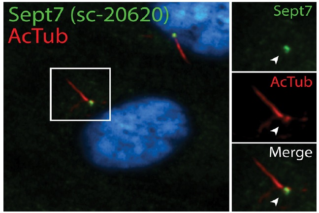
Septin 7 (H-120): sc-20620. Double immunofluorescent staining of methanol fixed human retinal pigment epithelial (RPE) cells using acetylated α Tubulin (6-11B-1): sc-23950 and donkey anti-mouse IgG-CFL 555: sc-362268 showing cilia staining (red immunofluorescence), Septin 7 (H-120): sc-20620 and donkey anti-rabbit IgG-FITC: sc-2090 showing base of cilia staining (green immunofluorescence) and Hoechst 33342: sc-391054 nuclear counterstain (blue immunofluorescence). Image kindly provided by Moshe Kim, Department of Cell Biology, Hospital for Sick Children (Toronto, Canada)
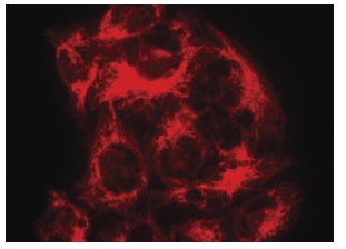
acetylated α Tubulin (6-11B-1): sc-23950. Immunofluorescence staining of methanol-fixed HeLa cells showing cytoplasmic localization.
acetylated α Tubulin (6-11B-1): sc-23950. Western blot analysis of acetylated α Tubulin in untreated (A) and Trichostatin A (sc-3511) treated (B) NIH/3T3 whole cell lysates. Note upregulation of acetylated α Tubulin in lane B.
Western blot analysis of acetylated α Tubulin acetylation in untreated (A) and Oxamflatin (sc-205960) treated (B) HeLa whole cell lysates. Antibodies tested include acetylated α Tubulin (6-11B-1): sc-23950 (A,B). Note acetylation of acetylated α Tubulin in lane B.
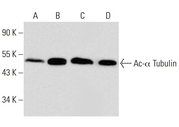
Western blot analysis of α Tubulin expression in untreated (A,C) and Panobinostat (sc-208148) treated (B,D) A549 whole cell lysates. Antibodies tested include acetylated α Tubulin (6-11B-1): sc-23950 (A,B) and α Tubulin (B-7): sc-5286 (C,D).
Western blot analysis of α Tubulin acetylation in untreated (A) and CBHA (sc-205240) treated (B) A549 whole cell lysates. Antibodies tested include acetylated α Tubulin (6-11B-1): sc-23950 (A,B) and α Tubulin (B-7): sc-5286 (C,D).
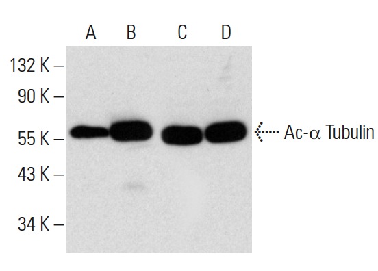
Western blot analysis of α Tubulin acetylation in untreated (A,C) and Scriptaid (sc-202807) treated (B,D) A549 whole cell lysates. Antibodies tested include acetylated α Tubulin (6-11B-1): sc-23950 (A,B) and α Tubulin (B-7): sc-5286 (C,D).
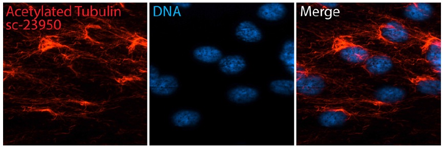
acetylated α Tubulin (6-11B-1): sc-23950. Immunofluorescent staining of methanol fixed human retinal pigment epithelial (RPE) cells showing microtubule-like staining (red immunofluorescence) and Hoechst 33342: sc-391054 nuclear counterstain (blue immunofluorescence). Secondary antibody used was donkey anti-mouse IgG-CFL 555: sc-362268 . Image kindly provided by Moshe Kim, Department of Cell Biology, Hospital for Sick Children (Toronto, Canada)
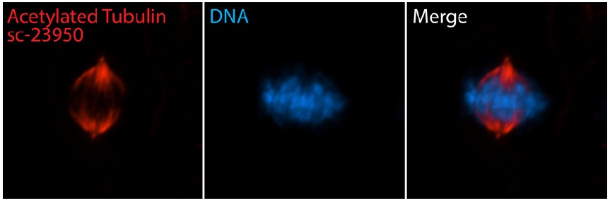
acetylated α Tubulin (6-11B-1): sc-23950. Immunofluorescent staining of methanol fixed human retinal pigment epithelial (RPE) cells showing mitotic spindle staining (red immunofluorescence) and Hoechst 33342: sc-391054 nuclear counterstain (blue immunofluorescence). Secondary antibody used was donkey anti-mouse IgG-CFL 555: sc-362268 . Image kindly provided by Moshe Kim, Department of Cell Biology, Hospital for Sick Children (Toronto, Canada)
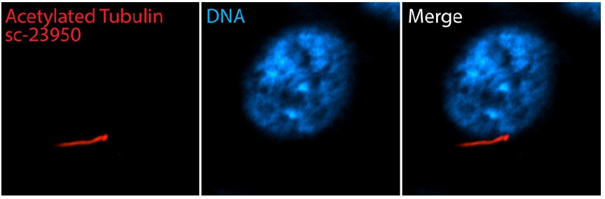
acetylated α Tubulin (6-11B-1): sc-23950. Immunofluorescent staining of methanol fixed, serum-starved human retinal pigment epithelial (RPE) cells showing cilia staining (red immunofluorescence) and Hoechst 33342: sc-391054 nuclear counterstain (blue immunofluorescence). Secondary antibody used was donkey anti-mouse IgG-CFL 555: sc-362268 . Image kindly provided by Moshe Kim, Department of Cell Biology, Hospital for Sick Children (Toronto, Canada)