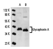
Glycophorin A (R10): sc-53905. Western blot analysis of Glycophorin A expression in K-562 (A) and human PBL (B) whole cell lysates.
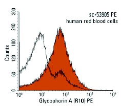
Glycophorin A (R10): sc-53905. FCM analysis of human red blood cells stained with Glycophorin A (R10) followed by PE-conjugated goat anti-mouse IgG: sc-3738. Black line histogram represents the isotype control, normal mouse IgG
1: sc-2855.
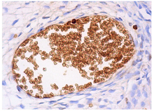
Glycophorin A (R10): sc-53905. Immunoperoxidase staining of formalin fixed, paraffin-embedded human blood vessel tissue showing membrane staining of red blood cells.