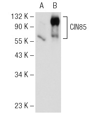
CIN85 (A-7): sc-166862. Western blot analysis of CIN85 expression in non-transfected: sc-117752 (A) and human CIN85 transfected: sc-175351 (B) 293T whole cell lysates.
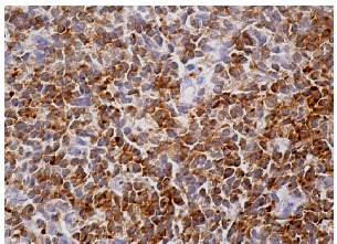
CIN85 (A-7): sc-166862. Immunoperoxidase staining of formalin fixed, paraffin-embedded human lymph node tissue showing cytoplasmic staining of cells in germinal and non-germinal centers.

CIN85 (A-7): sc-166862. Western blot analysis of CIN85 expression in THP-1 (A) and Jurkat (B) whole cell lysates.
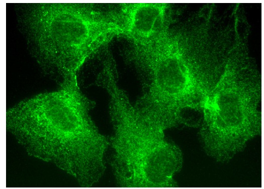
CIN85 (A-7): sc-166862. Immunofluorescence staining of formalin-fixed HepG2 cells showing cytoplasmic and membrane localization.