Bax (B-9): sc-7480. Immunoperoxidase staining of formalin-fixed, paraffin-embedded human breast carcinoma tissue showing membrane and cytoplasmic localization.
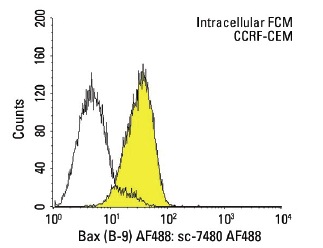
Bax (B-9) AF488: sc-7480 AF488. Intracellular FCM analysis of fixed and permeabilized CCRF-CEM cells. Black line histogram represents the isotype control, normal mouse IgG
2b: sc-3892.
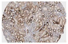
Bax (B-9): sc-7480. Immunoperoxidase staining of formalin fixed, paraffin-embedded human kidney tissue showing cytoplasmic staining of cells in glomeruli and tubuli. Kindly provided by The Swedish Human Protein Atlas (HPA) program.
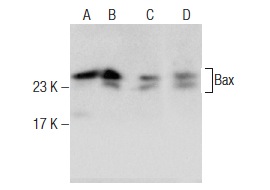
Western blot analysis of Bax expression in Jurkat (A), HuT 78 (B), Ramos (C) and BJAB (D) whole cell lysates. Antibodies tested include Bax (P-19): sc-526 (A,B) and Bax (B-9): sc-7480 (C,D).
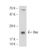
Bax (B-9): sc-7480. Western blot analysis of Bax expression in HuT 78 whole cell lysate.
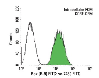
Bax (B-9) FITC: sc-7480 FITC. Intracellular FCM analysis of fixed and permeabilized CCRF-CEM cells. Black line histogram represents the isotype control, normal mouse IgG
2b: sc-2857.
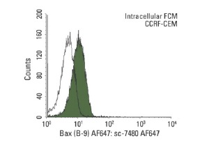
Bax (B-9) AF647: sc-7480 AF647. Intracellular FCM analysis of fixed and permeabilized CCRF-CEM cells. Black line histogram represents the isotype control, normal mouse IgG
2b: sc-24638.
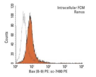
Bax (B-9) PE: sc-7480 PE. Intracellular FCM analysis of fixed and permeabilized Ramos cells. Black line histogram represents the isotype control, normal mouse IgG
2b: sc-2868.
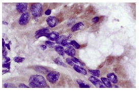
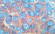
Bax (B-9): sc-7480. Immunoperoxidase staining of formalin fixed, paraffin-embedded human liver tumor showing cytoplasmic localization.
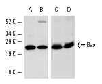
Bax (B-9): sc-7480. Western blot analysis of Bax expression in COLO 320DM (A), CCRF-CEM (B), HuT 78 (C) and BJAB (D) whole cell lysates.
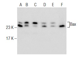
Bax (B-9): sc-7480. Western blot analysis of Bax expression in RAW 264.7 (A), CTLL-2 (B), Jurkat (C), MCF7 (D) and HT-1080 (E) whole cell lysates and mouse brain tissue extract (F).