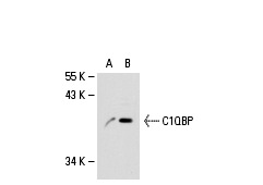
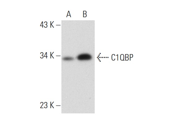
C1QBP (74.5.2): sc-23885. Western blot analysis of C1QBP expression in non-transfected: sc-110760 (A) and human C1QBP transfected: sc-113035 (B) 293 whole cell lysates.
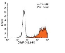
C1QBP (74.5.2) PE: sc-23885 PE. FCM analysis of human peripheral blood leukocytes. Black line histogram represents the isotype control, normal mouse IgG
1: sc-2866.
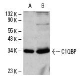
C1QBP (74.5.2): sc-23885. Western blot analysis of C1QBP expression in HL-60 (A) and Hep G2 (B) whole cell lysates.
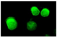
C1QBP (74.5.2): sc-23885. Immunofluorescence staining of methanol-fixed HL-60 cells showing cytoplasmic localization.
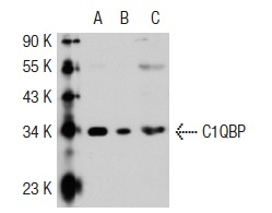
C1QBP (74.5.2): sc-23885. Western blot analysis of C1QBP expression in non-transfected 293T: sc-117752 (A), mouse C1QBP transfected 293T: sc-125076 (B) and Ramos (C) whole cell lysates.
C1QBP (74.5.2): sc-23885. Western blot analysis of C1QBP expression in non-transfected: sc-117752 (A) and human C1QBP transfected: sc-171633 (B) 293T whole cell lysates.
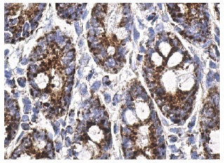
C1QBP (74.5.2): sc-23885. Immunoperoxidase staining of formalin fixed, paraffin-embedded human duodenum tissue showing cytoplasmic staining of glandular cells.