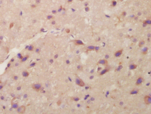
Formalin-fixed and paraffin embedded rat brain labeled with Anti-ITM2A Polyclonal Antibody, Unconjugated (bs-9705R) at 1:200 followed by conjugation to the secondary antibody and DAB staining\\n
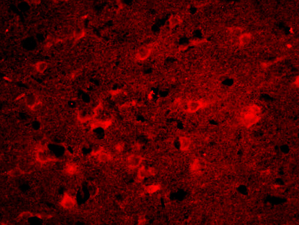
Formalin-fixed and paraffin-embedded rat brain labeled with Anti-ITM2A Polyclonal Antibody, Unconjugated(bs-9705R) 1:200, overnight at 4°C, The secondary antibody was Goat Anti-Rabbit IgG, Cy3 conjugated(bs-0295G-Cy3)used at 1:200 dilution for 40 minutes at 37°C.
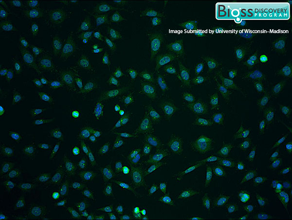
This image was kindly submitted by Dr. Lajoie from the University of Wisconsin-Madison.\\n4% PFA fixed rat brain vascular endothelial cells (RBE4) labeled with RABBIT ANTI-ITM2A POLYCLONAL ANTIBODY, UNCONJUGATED (BS-9705R) 1:100 dilution, followed by conjugated secondary antibody and DAPI staining. Anti-ITM2A staining is shown in green with blue DAPI counterstain.