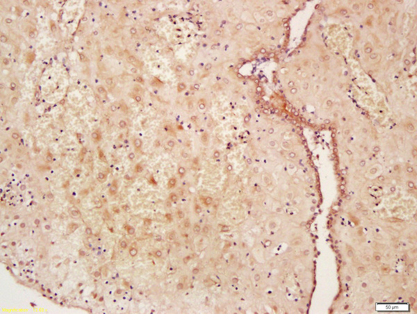
Formalin-fixed and paraffin embedded human placenta labeled with Anti-WNT2 Polyclonal Antibody, Unconjugated (bs-6133R) at 1:200 followed by conjugation to the secondary antibody and DAB staining\\n
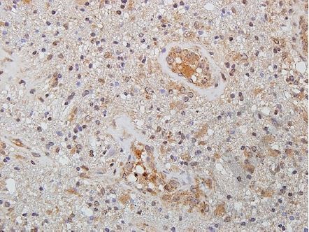
Images provided the Independent Validation Program (badge number 029629)Formalin-fixed and paraffin embedded human brain glioma tissue labeled with Rabbit Anti-WNT2 Polyclonal Antibody (bs-6133R) at 1:250 overnight at 4 °C followed by conjugation to secondary antibody.
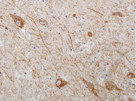
Images provided the Independent Validation Program (badge number 029629)Formalin-fixed and paraffin embedded human brain tissue with Parkinson's morphology labeled with Rabbit Anti-WNT2 Polyclonal Antibody (bs-6133R) at 1:250 overnight at 4 °C followed by conjugation to secondary antibody.
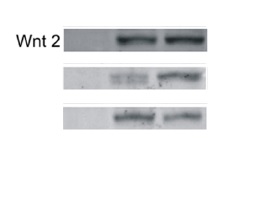
Image kindly provided by Dr. Magdalena Krol. Control tumor cells, tumor cells grown in macrophage-conditioned medium, tumor cells sorted from co-culture with macrophages, and macrophages from monocultures and sorted from co-culture with tumor cells were analyzed. Total protein concentrations in lysates were determined using a Bio-Rad protein assay. Proteins (50 mg) were resolved using SDS-PAGE and transferred onto PVDF membranes. The membranes were then blocked with 5% non-fat dry milk in TBS buffer containing 0.5% Tween 20. The membranes were then incubated overnight with the primary Rabbit Anti-WNT2 Polyclonal Antibody at 1:100 dilution. Subsequently, the membranes were washed three times in TBS containing 0.5% Tween 20 and incubated for 1 h at room temperature with secondary antibodies conjugated with the appropriate infrared (IR) fluorophore IRDyeH 800 CW or IRDyeH 680 RD at a dilution of 1:5000.
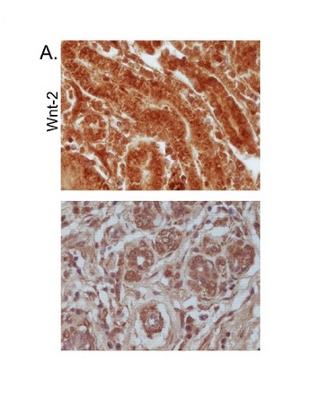
Image kindly submitted by Dr. Magdalena Krol. Formalin-fixed and paraffin embedded canine mammary tumor labeled with Rabbit Anti-WNT2 Polyclonal Antibody, Unconjugated (bs-6133R) at 1:200 followed by conjugation to the secondary antibody and DAB staining
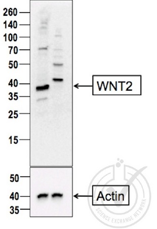
Image provided by the Independent Validation Program (badge number 29808). Lane 1: A549 cell extract, Lane 2: c6/36 mosquito cell extract (non-reactive\\nspecies) probed with Rabbit Anti-WNT2 Polyclonal Antibody, Unconjugated (bs-6133R) at 1:200 overnight at 4˚C. Followed by conjugation to secondary antibody at 1:10000 for 60 min at 26˚C.\\n
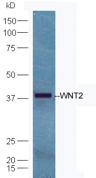
Mouse brain lysates probed with Anti-WNT2 Polyclonal Antibody, Unconjugated (bs-6133R) at 1:300 in 4˚C. Followed by conjugation to secondary antibody (bs-0295G-HRP) at 1:5000 90min in 37˚C.\\n


