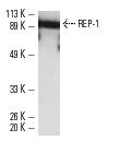
REP-1 (2F1): sc-23905. Western blot analysis of REP-1 expression in Y79 whole cell lysate.
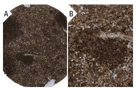
REP-1 (2F1): sc-23905. Immunoperoxidase staining of formalin fixed, paraffin-embedded human spleen tissue showing cytoplasmic staining of cells in red and white pulp (low and high magnification). Kindly provided by The Swedish Human Protein Atlas (HPA) program.
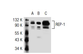
REP-1 (2F1): sc-23905. Western blot analysis of REP-1 expression in non-transfected 293T: sc-117752 (A), human REP-1 transfected 293T: sc-117225 (B) and Y79 (C) whole cell lysates.
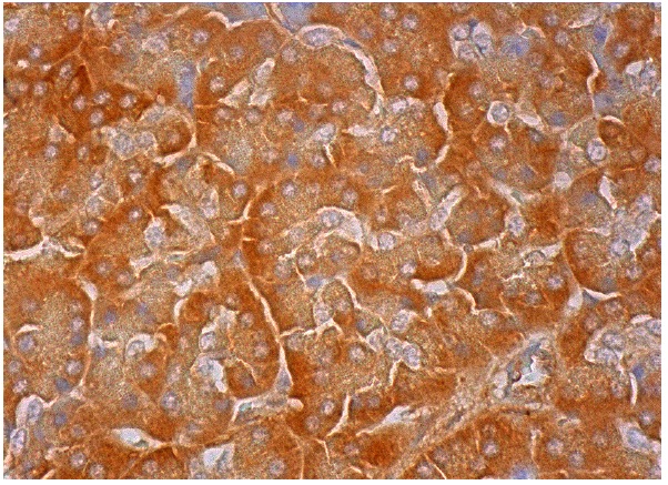
REP-1 (2F1): sc-23905. Immunoperoxidase staining of formalin fixed, paraffin-embedded human pancreas tissue showing cytoplasmic staining of exocrine glandular cells.