G
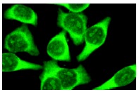
α q (E-17): sc-393. Immunofluorescence staining of methanol-fixed HeLa cells showing cytoplasmic staining.
G
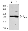
α q (E-17): sc-393. Western blot analysis of G
α q expression in Jurkat (A) and HeLa (B) whole cell lysates.
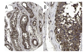
G
α q (E-17): sc-393. Immunoperoxidase staining of formalin fixed, paraffin-embedded human testis tissue showing cytoplasmic and membrane staining of cells in ductus seminiferous and Leydig cells at low (A) and high (B) magnification. Kindly provided by The Swedish Human Protein Atlas (HPA) program.
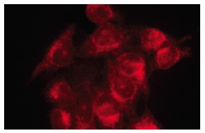
Gα q (E-17): sc-393. Immunofluorescence staining of methanol-fixed HeLa cells showing cytoplasmic localization.