S6 Ribosomal Protein (54D2) Mouse mAb (Alexa Fluor ® 488 Conjugate)
| Name | S6 Ribosomal Protein (54D2) Mouse mAb (Alexa Fluor ® 488 Conjugate) |
|---|---|
| Supplier | Cell Signaling Technology |
| Catalog | 5317 |
| Prices | $299.00 |
| Sizes | 100 µl (50 tests) |
| Host | Mouse |
| Clonality | Monoclonal |
| Isotype | IgG1 |
| Clone | 54D2 |
| Applications | ICC/IF FC |
| Species Reactivities | Human, Mouse, Rat, Monkey, Drosophila |
| Antigen | Monoclonal antibody is produced by immunizing animals with a recombinant fusion protein corresponding to full-length human S6 ribosomal protein. |
| Description | Mouse Monoclonal |
| Gene | RPS6 |
| Conjugate | Alexa Fluor ® 488 |
| Supplier Page | Shop |
Product images
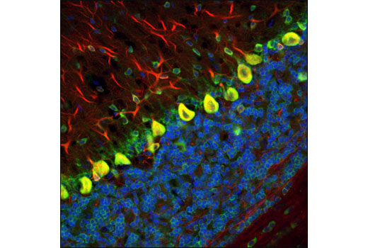 Confocal immunofluorescent analysis of rat brain using S6 Ribosomal Protein (54D2) (Alexa Fluor® 488) Mouse mAb (green) and β3-Tubulin (D71G9) XP ® Rabbit mAb #5568 (red). Blue pseudocolor = DRAQ5 ® (fluorescent DNA dye).
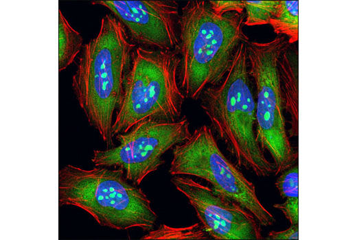 Confocal immunofluorescent analysis of HeLa cells using S6 Ribosomal Protein (54D2) Mouse mAb (Alexa Fluor ® 488 Conjugate) (green). Actin filaments were labeled with DY-554 phalloidin (red). Blue pseudocolor = DRAQ5 ® (fluorescent DNA dye).
 Flow cytometric analysis of Jurkat cells, using S6 Ribosomal Protein (54D2) Mouse mAb (Alexa Fluor® 488 Conjugate) (blue) compared to Mouse (MOPC-21) mAb IgG1 Isotype Control (Alexa Fluor® 488 Conjugate) #4878 (red).