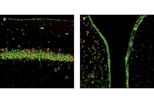
Confocal immunofluorescent analysis of rat brain showing the hippocampus (left) and striatum (right) labeled with Phospho-S6 Ribosomal Protein (Ser235/236) (2F9) Rabbit mAb (Alexa Fluor® 488 Conjugate) (green), EGR1 Antibody (red) #4152, and Phospho-Histone H3 (Ser10) (6G3) Mouse mAb #9706 (blue) following ischemia with 30 minute reperfusion.

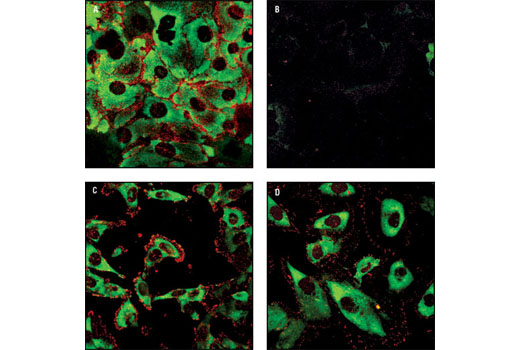
Confocal immunofluorescent analysis of two Gefinitib (Iressa) treated non-small cell lung cancer cell lines. HCC827 cells have the E746_A750 deletion in exon 19 of the EGFR gene, and are highly sensitive to Gefitinib. H1975 cells have the (T790M) mutation that confers Gefitinib-resistance. Both cell lines were treated and then double-labeled with Phospho-S6 Ribosomal Protein Rabbit mAb (Alexa Fluor® 488 Conjugate) #4854 and Phospho-Tyrosine Mouse mAb (Alexa Fluor® 647 Conjugate) #9415. Untreated HCC827 (A) and H1975 (C) cells show bright phospho-S6 (green) and phospho-tyrosine (red pseudocolor) label. Phospho-S6 and phospho-tyrosine signals dramatically decrease following Gefitinib treatment in HCC827 cells (B), with little or no change in the Gefitinib-resistant H1975 cells (D).

Flow cytometric analysis of Jurkat cells, untreated (green), or LY294002, Wortmannin and U0126-treated (blue), using Phospho-S6 Ribosomal Protein (Ser235/236) (2F9) Rabbit mAb (Alexa Fluor® 488 Conjugate) (#4854).


