Phospho-ATM/ATR Substrate (S*Q) (D23H2/D69H5) MultiMab™ Rabbit mAb mix
| Name | Phospho-ATM/ATR Substrate (S*Q) (D23H2/D69H5) MultiMab™ Rabbit mAb mix |
|---|---|
| Supplier | Cell Signaling Technology |
| Catalog | 9607 |
| Prices | $287.00 |
| Sizes | 100 µl (10 western blots) |
| Host | Rabbit |
| Clonality | Polyclonal |
| Isotype | IgG |
| Applications | WB IP |
| Antigen | MultiMab™ rabbit monoclonal mix antibodies are prepared by combining individual rabbit monoclonal clones in optimized ratios for the approved applications |
| Description | Rabbit Polyclonal |
| Gene | ATM |
| Supplier Page | Shop |
Product images
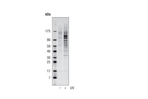 Western blot analysis of extracts from HeLa cells, untreated (-) or UV-treated (2 hr; +), using Phospho-ATM/ATR Substrate (S*Q) (D23H2/D69H5) MultiMab™ Rabbit mAb mix. Western blot was imaged using Odyssey ® Infrared Imaging System (LI-COR ® Biotechnologies).
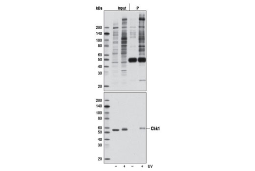 Immunoprecipitation of HeLa cells, untreated (-) or UV-treated (2 hr; +) (lanes 3 and 4), using Phospho-ATM/ATR Substrate (S*Q) (D23H2/D69H5) MultiMab™ Rabbit mAb mix. Lanes 1 and 2 are 10% input. Western blot analysis was performed using Phospho-ATM/ATR Substrate (S*Q) (D23H2/D69H5) MultiMab™ Rabbit mAb mix (upper) and Chk1 (2G1D5) Mouse mAb #2360 (lower).