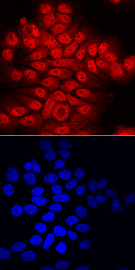
ATBF1/ZFHX3 was detected in immersion fixed MCF-7 human breast cancer cell line using Sheep Anti-Human ATBF1/ZFHX3 Antigen Affinity-purified Polyclonal Antibody (Catalog # AF7384) at 10 µg/mL for 3 hours at room temperature. Cells were stained using the NorthernLights™ 557-conjugated Anti-Sheep IgG Secondary Antibody (red, upper panel; Catalog # NL010) and counterstained with DAPI (blue, lower panel). Specific staining was localized to nuclei and cytoplasm. View our protocol for Fluorescent ICC Staining of Cells on Coverslips.