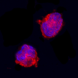
IL‑1ra/IL‑1F3 was detected in immersion fixed equine peripheral blood mononuclear cells (PBMCs) using Goat Anti-Equine IL‑1ra/IL‑1F3 Antigen Affinity-purified Polyclonal Antibody (Catalog # AF2466) at 15 µg/mL for 3 hours at room temperature. Cells were stained using the NorthernLights™ 557-conjugated Anti-Goat IgG Secondary Antibody (red; Catalog # NL001) and counterstained with DAPI (blue). Specific staining was localized to plasma membrane. View our protocol for Fluorescent ICC Staining of Non-adherent Cells.