Beta-1,3-N-Acetylglucosaminyltransferase 1/B3GNT1 Antibody
| Name | Beta-1,3-N-Acetylglucosaminyltransferase 1/B3GNT1 Antibody |
|---|---|
| Supplier | Novus Biologicals |
| Catalog | AF6664 |
| Prices | $99.00, $359.00 |
| Sizes | 25 µg, 100 µg |
| Host | Sheep |
| Clonality | Polyclonal |
| Isotype | IgG |
| Applications | WB |
| Species Reactivities | Human |
| Antigen | Chinese hamster ovary cell line CHO-derived recombinant human beta -1,3-N-acetylglucosaminyltransferase 1/B3GNT1 Asp43-Cys415 Accession # O43505 |
| Purity/Format | Immunogen affinity purified |
| Description | Sheep Polyclonal |
| Gene | B4GAT1 |
| Conjugate | Unconjugated |
| Supplier Page | Shop |
Product images
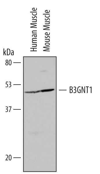 Western blot shows lysates of human muscle tissue and mouse muscle tissue. PVDF Membrane was probed with 1 µg/mL of Human beta -1,3-N-acetylglucosaminyltransferase 1/B3GNT1 Antigen Affinity-purified Polyclonal Antibody (Catalog # AF6664) followed by HRP-conjugated Anti-Sheep IgG Secondary Antibody (Catalog # HAF016). A specific band was detected for beta -1,3-N-acetylglucosaminyltransferase 1/B3GNT1 at approximately 50 kDa (as indicated). This experiment was conducted under reducing conditions and using Immunoblot Buffer Group 8.