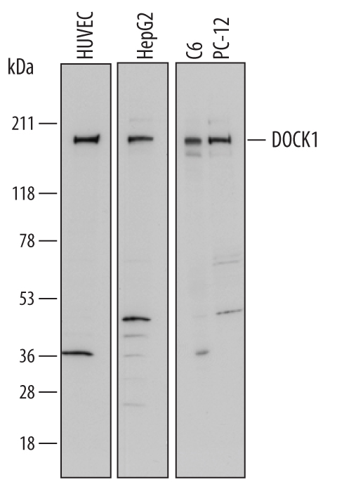
Western blot shows lysates of HUVEC human umbilical vein endothelial cells, HepG2 human hepatocellular carcinoma cell line, C6 rat glioma cell line, and PC‑12 rat adrenal pheochromocytoma cell line. PVDF membrane was probed with 1 µg/mL of Goat Anti-Human DOCK1 Antigen Affinity-purified Polyclonal Antibody (Catalog # AF7070) followed by HRP-conjugated Anti-Goat IgG Secondary Antibody (Catalog # HAF017). A specific band was detected for DOCK1 at approximately 180 kDa (as indicated). This experiment was conducted under reducing conditions and using Immunoblot Buffer Group 1.