
Western blot shows lysates of HepG2 human hepatocellular carcinoma cell line, Balb/3T3 mouse embryonic fibroblast cell line, and Rat-2 rat embryonic fibroblast cell line. PVDF membrane was probed with 1 µg/mL of Goat Anti-Human/Mouse/Rat APE Antigen Affinity-purified Polyclonal Antibody (Catalog # AF1044) followed by HRP‑conjugated Anti-Goat IgG Secondary Antibody (Catalog # HAF017). A specific band was detected for APE at approximately 40 kDa (as indicated). This experiment was conducted using Immunoblot Buffer Group 1.
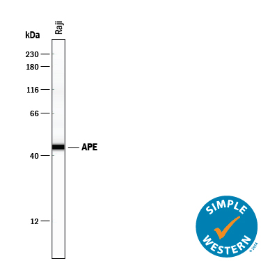
Simple Western lane view shows lysates of Raji human Burkitt's lymphoma cell line, loaded at 0.2 mg/mL. A specific band was detected for APE at approximately 45 kDa (as indicated) using 10 µg/mL of Goat Anti-Human/Mouse/Rat APE Antigen Affinity-purified Polyclonal Antibody (Catalog # AF1044) followed by 1:50 dilution of HRP-conjugated Anti-Goat IgG Secondary Antibody (Catalog # HAF109). This experiment was conducted under reducing conditions and using the 12-230 kDa separation system.