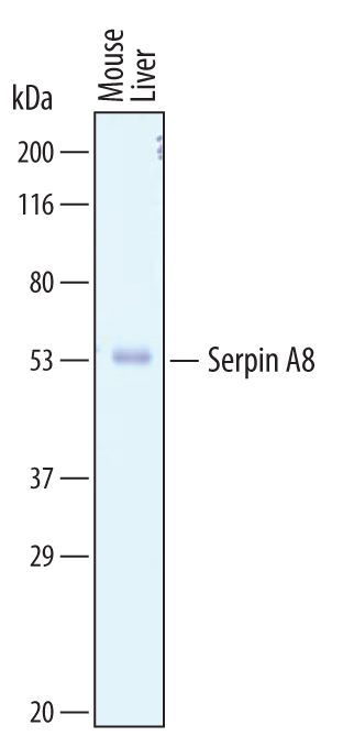
Western blot shows lysates of mouse liver tissue. PVDF membrane was probed with 1 µg/mL of Sheep Anti-Mouse Serpin A8/Angiotensinogen Antigen Affinity-purified Polyclonal Antibody (Catalog # AF6966) followed by HRP-conjugated Anti-Sheep IgG Secondary Antibody (Catalog # HAF016). A specific band was detected for Serpin A8/Angiotensinogen at approximately 55 kDa (as indicated). This experiment was conducted under reducing conditions and using Immunoblot Buffer Group 8.

Serpin A8/Angiotensinogen was detected in perfusion fixed frozen sections of mouse intestine using Sheep Anti-Mouse Serpin A8/Angiotensinogen Antigen Affinity-purified Polyclonal Antibody (Catalog # AF6966) at 1.7 µg/mL overnight at 4 °C. Tissue was stained using the Anti-Sheep HRP-DAB Cell & Tissue Staining Kit (brown; Catalog # CTS019) and counterstained with hematoxylin (blue). Specific staining was localized to the brush border in intestinal epithelium. View our protocol for Chromogenic IHC Staining of Frozen Tissue Sections.