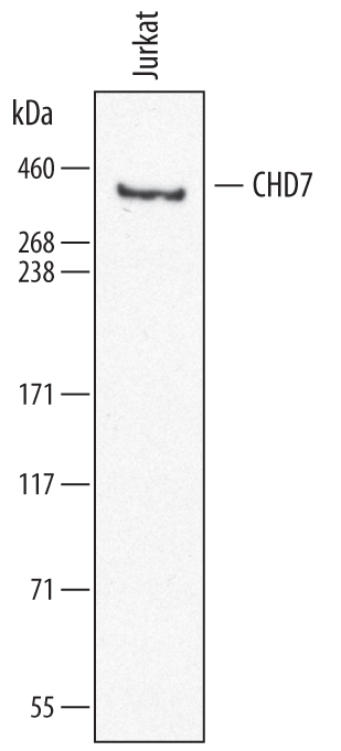
Western blot shows lysates of Jurkat human acute T cell leukemia cell line. PVDF membrane was probed with 0.5 µg/mL of Sheep Anti-Human CHD7 Antigen Affinity-purified Polyclonal Antibody (Catalog # AF7350) followed by HRP-conjugated Anti-Sheep IgG Secondary Antibody (Catalog # HAF016). A specific band was detected for CHD7 at approximately 350 kDa (as indicated). This experiment was conducted under reducing conditions and using Immunoblot Buffer Group 8.
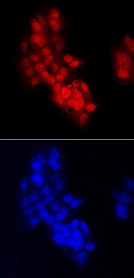
CHD7 was detected in immersion fixed HepG2 human hepatocellular carcinoma cell line using Sheep Anti-Human CHD7 Antigen Affinity-purified Polyclonal Antibody (Catalog # AF7350) at 10 µg/mL for 3 hours at room temperature. Cells were stained using the NorthernLights™ 557-conjugated Anti-Sheep IgG Secondary Antibody (red, upper panel; Catalog # NL010) and counterstained with DAPI (blue, lower panel). Specific staining was localized to nuclei. View our protocol for Fluorescent ICC Staining of Cells on Coverslips.